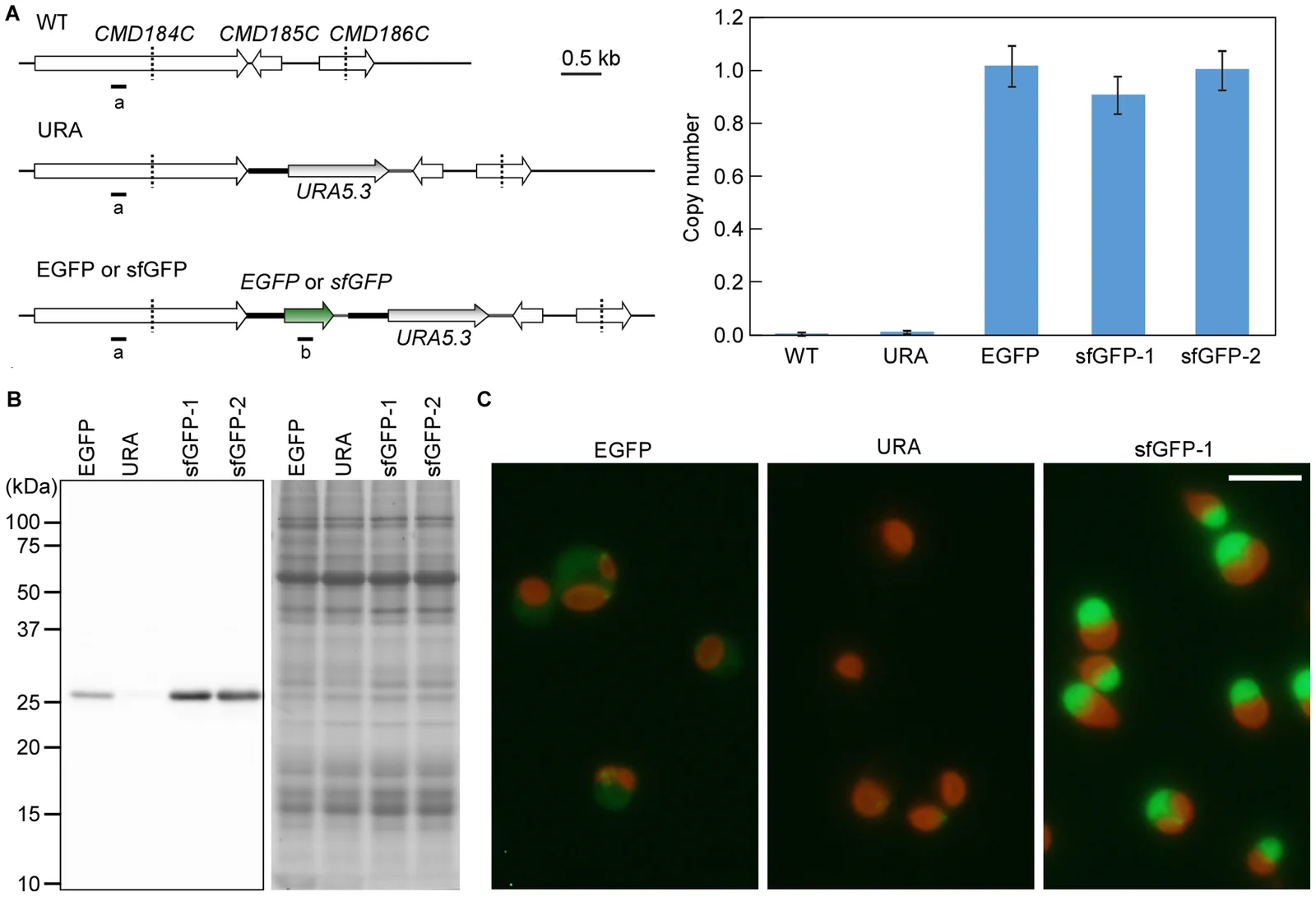
Fluorescence microscopy of experimental Cyanidioschyzon merolae transformants in panel C of Sumiya and colleagues’ 2014 figure 2, alongside genetic diagrams and protein measurements
Panel C shows Cyanidioschyzon merolae cells from experimental EGFP, URA and sfGFP-1 strains. Green GFP fluorescence and chlorophyll autofluorescence were captured together; these microscopy colours are not an ordinary daylight portrait. The scale bar is five micrometres. Panels A and B document introduced gene copies and protein measurements, rather than additional organisms.
Overview
Cyanidioschyzon merolae is a photosynthetic unicellular red alga studied for the coordination of cell and organelle division. Its cells normally contain one nucleus, one mitochondrion and one chloroplast. A light–dark cycle can synchronize division, allowing researchers to follow events that would otherwise occur at different times in different cells. The 2014 study by Sumiya and colleagues places it among the thermoacidophilic Cyanidiales, algae associated with unusually acidic, warm environments. Environmental tolerance and a particular laboratory treatment are different measurements: the experiment concerns cultured cells rather than a survey of wild populations.
The researchers developed a way to turn selected introduced genes on with heat. They cultured strain 10D or stable transformants in Allen’s medium under continuous illumination and aeration. A promoter from CMJ101C, a gene encoding a small heat-shock protein, controlled a superfolder green fluorescent reporter. Comparing promoter lengths was important because some constructs produced background expression before induction. A 200-base-pair upstream region had less leakage at lower temperatures than longer alternatives. In the stable S-200 strain, a shift to 50°C produced detectable reporter protein after thirty minutes and the highest measured protein level after approximately an hour. This was an experimentally optimized switch, not a newly discovered natural fluorescent trait.
The molecular readouts also behaved differently over time. Reporter RNA decreased rapidly after heat treatment ended, while fluorescent protein remained detectable a day later. The authors suggested protein stability could explain that persistence. They measured RNA, protein and fluorescence separately, using quantitative or semi-quantitative PCR, immunoblotting and fluorescence microscopy. These methods make the distinction between transcription and a lingering protein signal especially important; a glowing cell does not necessarily indicate that the introduced gene is still being transcribed at the earlier rate.
To investigate chloroplast separation, the team induced a modified DRP5B protein carrying the K135A substitution in its GTP-binding region. Some cells developed paired daughter chloroplasts linked by a small protein-positive bridge, with daughter nuclei on opposite sides. The observations support interference with final chloroplast scission rather than initial constriction. Cell-cycle marker expression subsequently rose and fell after renewed illumination, suggesting progression could continue despite incomplete separation. The proposed physical obstruction of cytokinesis remains an interpretation of the altered cells. This manipulation provides evidence about division machinery in a defined experimental system; it does not describe the usual shape or life cycle of every unmodified individual.
Origins & earliest records
A living red-algal species represented here by strain 10D and derived laboratory transformants. The 2014 publication dates an experimental method, not the evolutionary origin of the species.
Evidence & interpretation
Sumiya and colleagues compared promoter constructs, transcript and protein measurements, and microscopy of inducibly modified cells. Final scission and cell-cycle effects are inferred from those perturbations; no independent replication or supplementary-primer analysis is claimed here.
Selected bibliography
Documented works and useful reading. This is not a list of every appearance.
- 01
Development of a Heat-Shock Inducible Gene Expression System in the Red Alga Cyanidioschyzon merolae
PLOS ONE 9(10): e111261, 22 October. Complete main report, Materials and Methods and textual figure captions; DOI 10.1371/journal.pone.0111261. CC BY 4.0.
Family, evolution & connections
Selected archaeplastid nuclear branching (Schön et al., 2021)
Eight selected terminals from the 67-taxon analysis in Figure 2. Palpitomonas bilix represents sampled Cryptista. One composite Picozoa OTU is retained exactly as labelled; other Picozoa assemblies and most eukaryotes are omitted.
Scroll sideways to see all branches. With a keyboard, focus the tree and use the arrow keys.
- Selected nuclear sample tree
- Selected Archaeplastida (bootstrap 93 / posterior 1)
- Selected red-algal, rhodelphid and picozoan branch (bootstrap 100 / posterior 1)
- Selected red algae / rhodelphid branch (bootstrap 95 / posterior 1)
- Selected red-algal pair
- Galdieria sulphuraria
- Rhodelphis marinus
- COSAG02 / COSAG03 / COSAG04 / COSAG06 — composite source OTU
- Selected green-algal / glaucophyte branch
- Selected green lineage
- Volvox carteri
- Cyanophora paradoxa
- Palpitomonas bilix — selected cryptist outgroup
The selected tree uses 317 concatenated nuclear markers with LG + C60 + F + G-PMSF maximum likelihood and 100 non-parametric bootstrap replicates. Comparison posterior probabilities came from PhyloBayes CAT-GTR + G. Three original branch support pairs are retained, without recomputation after pruning. Branch lengths, remaining supports and the inset topology are omitted. Picozoa sister to red algae plus rhodelphids is the main Figure 2 result. An alternative joining Picozoa directly with red algae was also not rejected by the AU test and appeared after stronger site filtering. Some closely related sequences were merged into OTUs; Bayesian chains achieved only partial convergence. Nuclear branching does not establish which plastid-loss or independent-gain scenario occurred. Adapted from Schön et al. (2021), CC BY 4.0: eight tips retained, omitted branches suppressed and layout redrawn; three original support pairs retained.
Cyanidiococcus yangmingshanensis 8.1.23 F7
Cho and colleagues compared this genome with C. merolae 10D: nine chromosomes retained fully conserved gene order, while eleven showed divisions, fusions, inversions or relocations. This is a comparison of sampled genomes, not evidence that either living species is the other’s ancestor.
References
Sources supporting this profile. Linked pages have their own scope and editorial standards.
- ResearchDevelopment of a Heat-Shock Inducible Gene Expression System in the Red Alga Cyanidioschyzon merolaeResearch access: 2026-10-05
Image credits
Scientific organism fluorescence microscopy and experiment figureFluorescence microscopy of experimental Cyanidioschyzon merolae transformants in panel C of Sumiya and colleagues’ 2014 figure 2, alongside genetic diagrams and protein measurements
Panel C shows Cyanidioschyzon merolae cells from experimental EGFP, URA and sfGFP-1 strains. Green GFP fluorescence and chlorophyll autofluorescence were captured together; these microscopy colours are not an ordinary daylight portrait. The scale bar is five micrometres. Panels A and B document introduced gene copies and protein measurements, rather than additional organisms.