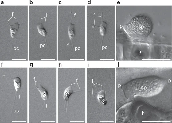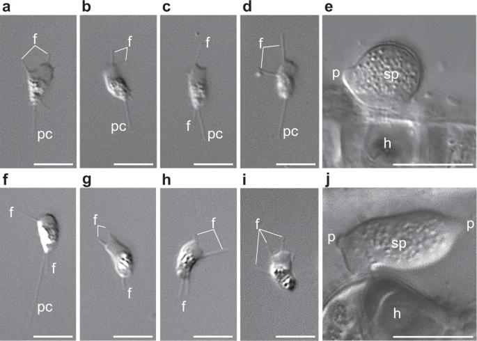
Sanchytrium tribonematis cells in upper panels a–e, compared with Amoeboradix gromovi in lower panels f–j
The upper row, panels a–e, shows Sanchytrium tribonematis: crawling zoospores and a sporangium on its host. The lower row belongs to the distinct species Amoeboradix gromovi. Letters mark filopodia, the posterior pseudocilium, host and sporangium. Scale bars differ between the motile cells and sporangia. The complete published microscopy figure is reproduced unchanged.
Overview
Sanchytrium tribonematis is a microscopic parasite of freshwater algae. Its spore-producing body sits on the outside of a host cell, while a fine, slightly branched rhizoid penetrates the host. The 2021 taxonomic account describes smooth, rounded or oval sporangia about ten micrometres in diameter, usually with no visible discharge papilla or a single one. The cellular stages emerging from these structures are amoeboid: they move over surfaces using an anterior sheet-like protrusion and finer projections extending laterally and behind.
A trailing structure resembles a flagellum but does not propel the cell through water. The authors call it a pseudocilium. In this species its supporting basal body consists of nine single microtubules, rather than the triplets typical of many other flagellated eukaryotes. The trailing part can reach roughly five micrometres. These details distinguish a reduced structure from an ordinary swimming flagellum; the cells examined in these studies were observed crawling, not swimming.
Galindo and colleagues sequenced DNA from an individually isolated sporangium of culture strain X-128 after amplifying its DNA. Because the organism was maintained with the yellow-green alga Tribonema gayanum, the researchers screened the resulting sequences for both bacterial and host contamination. Their final assembly was about 11.2 million DNA bases. Roughly half of the predicted proteins could receive a functional annotation, so an unrecognized sequence should not automatically be treated as a gene with no biological function.
The combined sequence analyses place Sanchytrium with Amoeboradix gromovi in the newly proposed phylum Sanchytriomycota, as a sister lineage to Blastocladiomycota. Two conserved-protein datasets support this relationship. The paper also investigates how rapidly evolving sequences can mislead deeper fungal branching, testing different taxon selections and removal of fast-changing sites. The classification represents that comparative analysis, rather than a visible property inferred from a photograph.
The retained pseudocilium may have an additional role. Both sanchytrids possess candidate proteins related to a light-sensing system studied in another fungus, and their reduced flagellar apparatus might help support a sensory structure. Functional experiments are still needed to establish this proposed role. The species therefore illustrates a combination of observed crawling behavior, reduced cellular architecture and testable genomic hypotheses, without being described as a fungus that can see images.
Origins & earliest records
Galindo and colleagues analyzed culture strain X-128 in 2021 and provided an updated taxonomic appendix. The strain was maintained with Tribonema gayanum strain 20 CALU. This account does not designate the sequenced culture as a holotype or independently inspect the earlier original species description.
Evidence & interpretation
The primary article combines light microscopy, single-sporangium genome sequencing, contamination screening and conserved-protein phylogenies. Anatomy and crawling behavior are distinguished from the proposed sensory role of the residual flagellar structure.
Selected bibliography
Documented works and useful reading. This is not a list of every appearance.
- 01
Phylogenomics of a new fungal phylum reveals multiple waves of reductive evolution across Holomycota
Nature Communications 12,4973; complete relevant phylogenetic, flagellar and sensory Results/Discussion, taxonomic appendix, biological-material, sequencing, assembly and phylogenomic Methods. Original adaptation of CC BY 4.0 article. https://creativecommons.org/licenses/by/4.0/.
Family, evolution & connections
Sanchytrids and sampled blastocladiomycetes — Galindo 2021
Five species retained from Figure 2a’s Sanchytriomycota–Blastocladiomycota clade. Other fungi and outgroups omitted.
Scroll sideways to see all branches. With a keyboard, focus the tree and use the arrow keys.
- Selected clade (posterior ≥0.99; ML bootstrap ≥99%)
- Sanchytriomycota (posterior ≥0.99; ML bootstrap ≥99%)
- Sampled Blastocladiomycota (posterior ≥0.99; ML bootstrap ≥99%)
- Allomyces macrogynus
- Blastocladiella / Catenaria pair (posterior ≥0.99; ML bootstrap ≥99%)
- Blastocladiella emersonii
- Catenaria anguillulae
GBE59 analysis: 264 conserved proteins, 93,743 amino-acid positions and 59 species. Bayesian CAT-Poisson analysis and maximum-likelihood PMSF analysis agree on this clade. Black-dot thresholds are retained from the original figure, not recalculated. Fast-evolving sanchytrids can distort deeper fungal rooting; alternative taxon sets and site removal were tested. This subtree makes no universal fungal-root claim. The separate 53-protein BMC analysis is not blended into these node labels. No dates, direct ancestors or species ages inferred.
Taxonomic classification
A hierarchy of classified groups, not a chain of direct ancestors.
- KingdomFungi
- PhylumSanchytriomycota
- GenusSanchytrium
- SpeciesSanchytrium tribonematis
Amoeboradix gromovi
Both species form a supported clade in the 2021 protein analyses, proposed as Sanchytriomycota. Their long branches reflect rapid sequence evolution, not greater age or direct ancestry between them.
Sampled Blastocladiomycota
The sampled blastocladiomycetes form the sister branch to sanchytrids. This relationship survives the study’s long-branch and fast-site tests, while deeper rooting among major fungal groups remains more sensitive to analysis choices.
Tribonema gayanum
The 2021 genome study maintained Sanchytrium strain X-128 on this freshwater yellow-green alga and collected spores and sporangia after infection. This identifies an experimental host association; it does not establish an exhaustive natural host range.
References
Sources supporting this profile. Linked pages have their own scope and editorial standards.
- ResearchGalindo et al. 2021 primary comparative study and taxonomic appendixResearch access: 2026-10-05
Image credits
Scientific organism microscopy photographSanchytrium tribonematis cells in upper panels a–e, compared with Amoeboradix gromovi in lower panels f–j
The upper row, panels a–e, shows Sanchytrium tribonematis: crawling zoospores and a sporangium on its host. The lower row belongs to the distinct species Amoeboradix gromovi. Letters mark filopodia, the posterior pseudocilium, host and sporangium. Scale bars differ between the motile cells and sporangia. The complete published microscopy figure is reproduced unchanged.