A sourced illustration has not yet been added. This gap is included in the coverage record.
Overview
Potteromyces asteroxylicola grew in the outer tissues of Asteroxylon mackiei, a small lycopsid plant from the Rhynie chert. Its thread-like hyphae formed a compact mound beneath the plant surface, while clusters of spore-bearing stalks pushed through the cuticle. Strullu-Derrien and colleagues describe stalks approximately 150–300 micrometres long and 7.5–11.5 micrometres wide, with rounded conidia 6.5–10.5 micrometres across. These are asexual spores; no sexual reproductive body has been identified for this species.
The surrounding plant tissue makes this fossil particularly informative. Dome-shaped projections contain differently shaped cells alongside fungal filaments. The authors interpret this local proliferation as a response by the living plant to infection, rather than a fungus simply feeding on tissues after death. Other tissue systems show decay before petrification, but the localized host reaction supports an earlier disease process. The study proposes a necrotrophic lifestyle, in which a pathogen kills host tissue before obtaining nutrition from it. That interpretation does not identify a modern crop disease or prove that every Rhynie fungus was harmful.
Two historical rock sections contain the evidence. Ordinary transmitted-light microscopy documents the overall association, while confocal microscopy combines many optical slices into views of deeper structures. Some displayed colors encode depth and are not original fungal colors. Rounded spores are visible near the stalk tips and sometimes in chains; nearby fallen spores cannot always be connected physically with a particular stalk. The fine details of spore formation therefore remain qualified in the description.
The researchers distinguish Potteromyces from the previously described Paleopyrenomycites devonicus by the shape and dimensions of its stalks and spores, and its occurrence on both aerial axes and leaf-like outgrowths. Superficially similar structures occur among living ascomycetes, but none gives a secure placement in a modern class or order. The authors favor an extinct ascomycete lineage of uncertain position. They specifically caution that incorrectly assigning an ancient fossil to a narrow living group can distort molecular-clock estimates.
The genus honors Beatrix Potter’s work as a naturalist and observer of fungi. Its recognition in 2023 came from exceptionally preserved Early Devonian material, including slides prepared roughly a century earlier. The discovery documents a particular ancient plant–fungus interaction; it does not establish the first pathogen that ever evolved or make this species the direct ancestor of living pathogenic fungi.
Origins & earliest records
Strullu-Derrien and Hawksworth named the genus and species in 2023. Holotype NHMUK-V 16430 is a thin section prepared around 1915 by W.Hemingway; isotype NMS G.1925-9-11 is in National Museums Scotland. The study attributes the Rhynie chert a radiometric age of 407.1±2.2 Ma.
Evidence & interpretation
The 2023 primary study documents fungal stalks, conidia, hyphae and host reaction tissues using white-light and confocal microscopy. Figures 1–2 show the holotype and Figures 3–4 the isotype. Infection before host death is inferred from the localized tissue reaction; finer fungal relationships remain unresolved. Image observations are not replicated infection experiments.
Selected bibliography
Documented works and useful reading. This is not a list of every appearance.
- 01
A fungal plant pathogen discovered in the Devonian Rhynie Chert
Nature Communications14,7932; complete main Results, taxonomy, Discussion and Methods. Original adaptation of CC BY4.0 article; https://creativecommons.org/licenses/by/4.0/.
Family, evolution & connections
Taxonomic classification
A hierarchy of classified groups, not a chain of direct ancestors.
- KingdomFungi
- PhylumAscomycota
- GenusPotteromyces
- SpeciesPotteromyces asteroxylicola
Asteroxylon mackiei
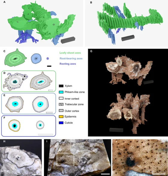
Fungal structures penetrate the plant cuticle alongside localized tissue reactions. The authors interpret this association as infection of a living host and probable necrotrophic pathogenicity; fossil anatomy does not directly record feeding behavior.
References
Sources supporting this profile. Linked pages have their own scope and editorial standards.
- ResearchStrullu-Derrien et al.2023 primary descriptionResearch access: 2026-10-05
Image credits
A sourced illustration has not yet been added. This gap is included in the coverage record.