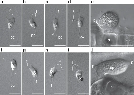
Amoeboradix gromovi cells in lower panels f–j, compared with Sanchytrium tribonematis in upper panels a–e
The lower row, panels f–j, shows Amoeboradix gromovi: crawling zoospores, including cells with a retracted pseudocilium, and a sporangium on its host. The upper row belongs to the distinct species Sanchytrium tribonematis. The annotations and separate five- and ten-micrometre scale bars are retained. The complete published microscopy figure is reproduced unchanged.
Overview
Amoeboradix gromovi is a small parasitic fungus whose active dispersal cells crawl rather than swim. They have broad and narrow cellular projections and a trailing pseudocilium, a reduced flagellar structure that can retract completely into the cell. The 2021 taxonomic appendix describes cells about two to three by three to four micrometres, with a pseudocilium reaching eight micrometres. Its exceptionally long basal body, approximately 1.8–2.2 micrometres, contains simplified microtubule singlets or doublets.
The spore-producing structures differ from those of its close relative Sanchytrium. Amoeboradix sporangia range from pear-shaped bodies to curved, asymmetrical sacs, with a conspicuous rhizoid and two or three discharge papillae. The reported sac-like form is roughly 8–10 micrometres wide and 16–18 micrometres long. The account also identifies small spherical cysts and larger, thick-walled resting spores. These are different stages of the fungus, not a single body repeatedly changing into unrelated organisms.
The genomic investigation cultured strain X-113 on Tribonema gayanum, a freshwater yellow-green alga. Researchers separated a mature sporangium from its host with a microscopic blade, washed it and amplified its DNA for sequencing. Removing bacterial and algal contamination was important because the parasite and its food source occurred together. The resulting fungal assembly contained approximately 10.5 million DNA bases and predicted 7,220 proteins; those values identify this study’s assembly rather than a complete census of variation throughout the species.
Comparisons with other fungi support a close relationship to Sanchytrium tribonematis. The authors place their shared lineage in Sanchytriomycota, sister to Blastocladiomycota, using two conserved-protein datasets and tests addressing unusually fast sequence evolution. Their genomes are reduced relative to several sampled zoosporic fungi, yet retain core mitochondrial machinery. Reduction therefore does not imply loss of all respiration or a universal tendency for parasites to become the same kind of organism.
A chain of lipid bodies near the long basal body provides a further research question. Together with candidate light-response proteins, it suggests that the residual flagellar apparatus could support a sensory function after losing its role in propulsion. The paper explicitly treats this as a hypothesis requiring functional study. Its current evidential strength lies in the observed cell architecture and molecular comparisons, not a demonstrated visual organ or a reconstructed direct ancestor of other fungi.
Origins & earliest records
Galindo and colleagues analyzed culture strain X-113 in 2021 and provided an updated taxonomic appendix. The strain was maintained with Tribonema gayanum strain 20 CALU. This account does not designate the sequenced culture as a holotype or independently inspect the earlier original species description.
Evidence & interpretation
The primary article combines light microscopy, single-sporangium genome sequencing, contamination screening and conserved-protein phylogenies. Anatomy and crawling behavior are distinguished from the proposed sensory role of the residual flagellar structure.
Selected bibliography
Documented works and useful reading. This is not a list of every appearance.
- 01
Phylogenomics of a new fungal phylum reveals multiple waves of reductive evolution across Holomycota
Nature Communications 12,4973; complete relevant phylogenetic, flagellar and sensory Results/Discussion, taxonomic appendix, biological-material, sequencing, assembly and phylogenomic Methods. Original adaptation of CC BY 4.0 article. https://creativecommons.org/licenses/by/4.0/.
Family, evolution & connections
Sanchytrids and sampled blastocladiomycetes — Galindo 2021
Five species retained from Figure 2a’s Sanchytriomycota–Blastocladiomycota clade. Other fungi and outgroups omitted.
Scroll sideways to see all branches. With a keyboard, focus the tree and use the arrow keys.
- Selected clade (posterior ≥0.99; ML bootstrap ≥99%)
- Sanchytriomycota (posterior ≥0.99; ML bootstrap ≥99%)
- Sampled Blastocladiomycota (posterior ≥0.99; ML bootstrap ≥99%)
- Allomyces macrogynus
- Blastocladiella / Catenaria pair (posterior ≥0.99; ML bootstrap ≥99%)
- Blastocladiella emersonii
- Catenaria anguillulae
GBE59 analysis: 264 conserved proteins, 93,743 amino-acid positions and 59 species. Bayesian CAT-Poisson analysis and maximum-likelihood PMSF analysis agree on this clade. Black-dot thresholds are retained from the original figure, not recalculated. Fast-evolving sanchytrids can distort deeper fungal rooting; alternative taxon sets and site removal were tested. This subtree makes no universal fungal-root claim. The separate 53-protein BMC analysis is not blended into these node labels. No dates, direct ancestors or species ages inferred.
Taxonomic classification
A hierarchy of classified groups, not a chain of direct ancestors.
- KingdomFungi
- PhylumSanchytriomycota
- GenusAmoeboradix
- SpeciesAmoeboradix gromovi
References
Sources supporting this profile. Linked pages have their own scope and editorial standards.
- ResearchGalindo et al. 2021 primary comparative study and taxonomic appendixResearch access: 2026-10-05
Image credits
Scientific organism microscopy photographAmoeboradix gromovi cells in lower panels f–j, compared with Sanchytrium tribonematis in upper panels a–e
The lower row, panels f–j, shows Amoeboradix gromovi: crawling zoospores, including cells with a retracted pseudocilium, and a sporangium on its host. The upper row belongs to the distinct species Sanchytrium tribonematis. The annotations and separate five- and ten-micrometre scale bars are retained. The complete published microscopy figure is reproduced unchanged.