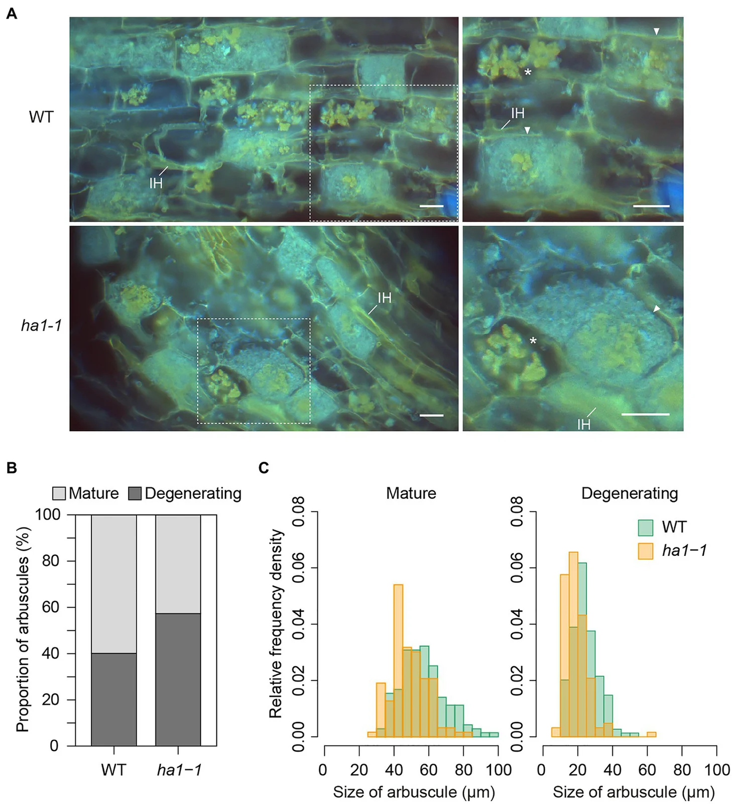
DAPI-stained Rhizophagus irregularis arbuscules in wild-type and ha1-1 Lotus japonicus roots, with Nguyen and Saito’s 2021 measurements
The upper panels show fungal arbuscules and intraradical hyphae inside Lotus japonicus roots four weeks after inoculation with Rhizophagus irregularis DAOM 197198. They compare wild-type and ha1-1 plant roots. Yellow and blue fluorescence arise from DAPI staining, rather than natural fungal colours. The lower panels quantify mature and degenerating arbuscules in these experimental conditions.
Overview
Rhizophagus irregularis is an arbuscular mycorrhizal fungus whose threads grow beyond plant roots into the surrounding substrate. The fungus is multinucleate, and its partnership with plants involves access to minerals and plant-derived organic nutrients. This organism is a model for investigating how a root-associated symbiont maintains and organizes its genome. Bethan F. Manley and colleagues assembled the laboratory isolate DAOM197198 in 2023, combining long DNA reads with chromosome-contact data and manual curation. The sequenced material was commercial spores of that isolate, rather than a newly designated taxonomic type or a survey of wild populations.
The resulting nuclear assembly contained approximately 146.8 million base pairs, arranged into 32 chromosome-scale scaffolds with ten remaining gaps. Twenty-three scaffolds were gapless, and seventeen carried identified telomeric sequence at both ends. Those are assembly characteristics, not an independent microscopic count of the fungus's chromosomes. The authors suggested that a joining error could explain why an earlier assembly had 33 scaffolds. They also reconstructed a separate, circular mitochondrial genome of 70,793 base pairs. The improved nuclear reference therefore clarified continuity and sequence organization while leaving some chromosome-structure questions open.
Short and long RNA reads helped refine 30,209 predicted gene models, including exon boundaries, untranslated regions and alternative transcripts. The long-read evidence corrected structures that earlier annotations had misrepresented. Comparative searches placed many nutrient-transport functions before the emergence of the Glomeromycotina, while also identifying genes inferred to be restricted to that lineage. Gene ages were analytical estimates, with a specific test for failure to detect distant homologues; they were not directly dated birth events. The inferred loss of a fatty-acid-synthase gene was discussed as part of the lineage's dependence on externally supplied lipids.
Across the assembled chromosomes, regions containing more conserved genes tended to show abundant long transcripts, whereas regions around evolutionarily younger genes produced more small RNAs. Importantly, the association did not mean that young genes themselves necessarily generated those small RNAs. The authors compared this organization with compartmentalized genomes in other fungi but explicitly lacked quantitative evidence for different evolutionary speeds between these regions. The reference thus offers testable patterns of genome organization, not proof that every proposed compartment has an established adaptive function.
A separate 2025 experiment used isolate MUCL43194, Rahnella aquatilis HX2 and Medicago root cultures. Twenty-four hours after bacterial addition, low-phosphorus conditions strengthened organic-phosphorus consumption, phosphatase activity and bacterial abundance in the joint treatment. Transcript changes supported altered nutrient cooperation, but they did not directly measure plant-to-fungus carbon flux. This controlled, two-compartment system distinguishes early microbial responses from field performance or a universal agricultural recommendation.
Origins & earliest records
Model arbuscular mycorrhizal fungus; the 2023 reference represents isolate DAOM197198.
Evidence & interpretation
Long-read assembly, chromosome-contact curation and transcript annotation support the genomic account; evolutionary gene ages and functional compartments remain inferred.
Selected bibliography
Documented works and useful reading. This is not a list of every appearance.
- 01
A highly contiguous genome assembly reveals sources of genomic novelty in the symbiotic fungus Rhizophagus irregularis
G3 13:jkad077, DOI 10.1093/g3journal/jkad077; complete main report and Methods. CC BY 4.0.
- 02
Early-stage reciprocal cooperation between the arbuscular mycorrhizal fungus Rhizophagus irregularis and the phosphate-solubilizing bacterium Rahnella aquatilis is dependent on external phosphorus availability
Communications Biology 8:1075; controlled early-stage experiment. CC BY-NC-ND 4.0.
- 03
Genome of an arbuscular mycorrhizal fungus provides insight into the oldest plant symbiosis
PNAS110:20117–20122;25Novemberonline,10Decemberissue.
Family, evolution & connections
Taxonomic classification
A hierarchy of classified groups, not a chain of direct ancestors.
- kingdomFungi
- subphylumGlomeromycotina
- genusRhizophagus
- speciesRhizophagus irregularis
Rahnella aquatilis HX2
Duan’s in-vitro model found stronger carbon–phosphorus exchange under low phosphorus; field communities remain untested.
Located primary passages
Specific passages supporting details in this entry, grouped by their published witness.
Primary source witness
2013 paper with2014 correction.
- For DAOM-197198, the assembled sequence covered101Mb against an estimated153Mb genome. Repetitive regions help explain that gap. This reference is a sampled reconstruction, not a complete sequence of every nucleus or isolate.
Complete Introduction20117–20118 and Genome Assembly20118; attached author correction563.
Remaining Results/Discussion, Methods and supplements uninspected. Native403/API500 and laterreader403 held; no localPDF/pixel claim.
References
Sources supporting this profile. Linked pages have their own scope and editorial standards.
- ResearchManley and colleagues 2023, official Europe PMC full text, CC BY 4.0Research access: 2026-10-05
- ResearchDuan and colleagues 2025, primary experimentResearch access: 2026-10-05
Image credits
Scientific fungal fluorescence microscopy and experiment figureDAPI-stained Rhizophagus irregularis arbuscules in wild-type and ha1-1 Lotus japonicus roots, with Nguyen and Saito’s 2021 measurements
The upper panels show fungal arbuscules and intraradical hyphae inside Lotus japonicus roots four weeks after inoculation with Rhizophagus irregularis DAOM 197198. They compare wild-type and ha1-1 plant roots. Yellow and blue fluorescence arise from DAPI staining, rather than natural fungal colours. The lower panels quantify mature and degenerating arbuscules in these experimental conditions.