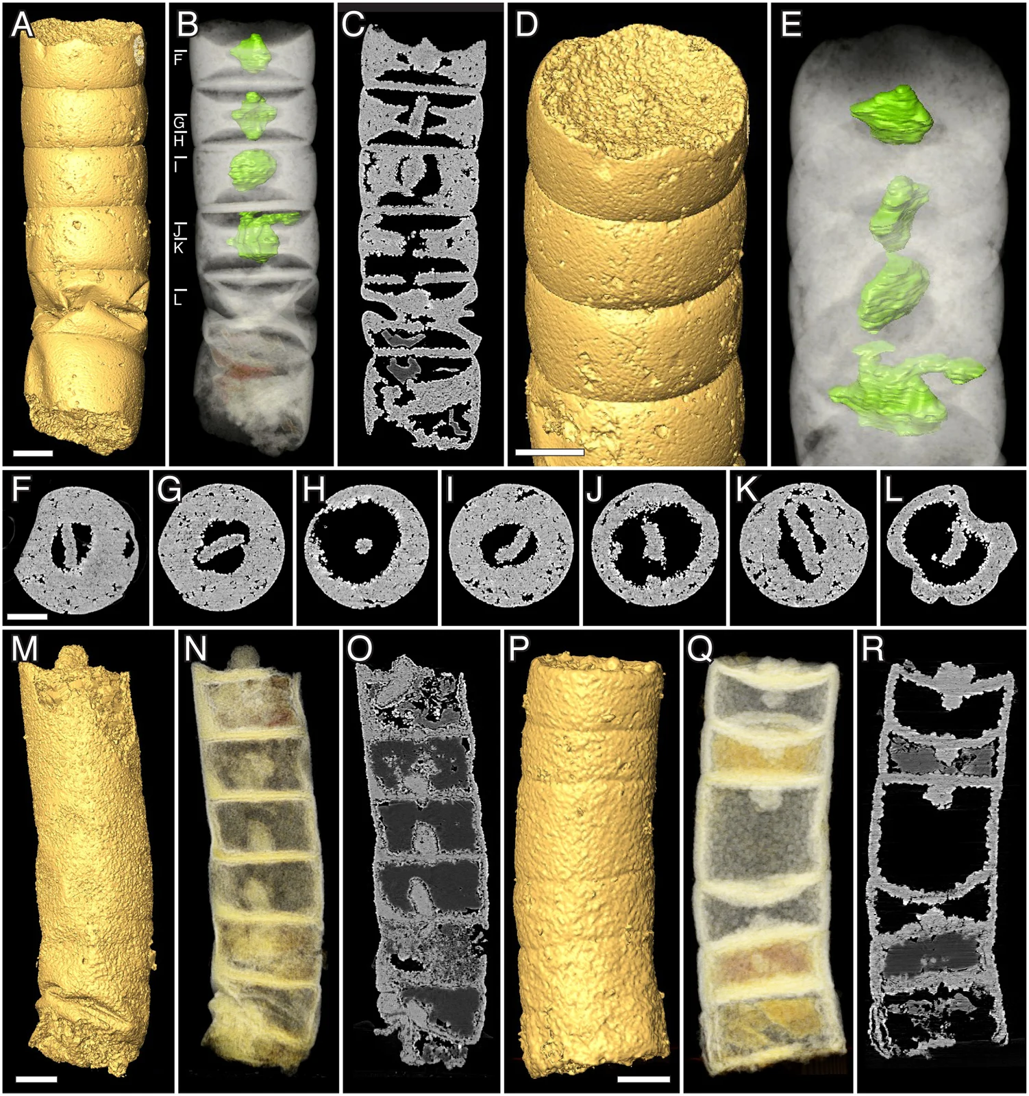
Synchrotron tomographic renderings and sections of Rafatazmia chitrakootensis fossils, including holotype NRM X4258
Panels A–L show holotype NRM X4258; the remaining panels show two further specimens. Surface renderings, transparent volumes and virtual sections reveal preserved internal structures. Green highlights were added for visibility and do not establish the organism’s living colour. Scale bars represent 50 micrometres. The published figure is reproduced unchanged.
Overview
Rafatazmia chitrakootensis consists of an unbranched row of compartments separated by transverse walls. Bengtson and colleagues measured tubes 58–175 micrometres wide and preserved fragments up to roughly 2 mm long. Neither original end is preserved, so fragment length is not necessarily the organism’s full length. Different neighboring compartments can vary markedly in size. Some dividing walls remain incomplete, supporting growth through partitioning at several places along the filament instead of solely adding cells at a specialized tip.
The best-preserved specimen, holotype NRM X 4258, contains six compartments and striking flattened, diamond-shaped internal bodies. Five compartments clearly preserve these bodies, suspended near their centers by surrounding material. Their repeated position, shape and alignment distinguish them from irregular mineral fillings elsewhere in the fossils. The authors interpret them as possible pyrenoids: protein-rich structures associated with the photosynthetic machinery of many algae. This interpretation identifies a likely function, rather than recovering original proteins, pigments or a living chloroplast.
Cell boundaries provide another proposed clue. Central openings in developing partitions and small bodies associated with other partition centers resemble the connections and plugs found between cells of red algae. Mineral alteration prevents a definitive identification of those structures as red-algal pit plugs. A single centrally positioned pyrenoid also is not uniquely diagnostic of red algae. The paper consequently treats a red-algal affinity as probable and a more specific bangiophycean relationship as uncertain, with the possibility that diffuse filament growth is an older shared character.
These fossils were preserved within phosphate-bearing microbial deposits of the Tirohan Dolomite. Early fine-grained apatite could replace biological structures, while later crystalline layers coated walls and filled cavities. Recognizing that sequence is essential: not every object inside a compartment is an original organelle. Tomographic virtual sections allowed the researchers to examine internal structures without mistaking an external view for complete anatomy.
The 2017 study attributes an age of approximately 1.6 billion years to the deposit, comparing direct phosphorite dating with regional zircon dates and stratigraphic correlations. If both that chronology and the proposed identity are correct, the fossil records very early multicellular red algae. This conditional conclusion does not establish Rafatazmia as a direct ancestor of living algae, or prove that its inferred photosynthetic structures are unambiguously identified.
Origins & earliest records
Named in 2017 by Bengtson, honoring Rafat Azmi; the species name refers to Chitrakoot. Type stratum is the Tirohan Dolomite at Jankikund. The approximate 1,600 Ma timeline follows the primary study’s geological interpretation, not a date measured from an individual filament.
Evidence & interpretation
Holotype Swedish Museum of Natural History NRM X 4258 appears in Figure 5 A–L. Figures 2–9 distinguish microscopy, tomographic renderings and virtual slices. The study discusses direct phosphorite Pb–Pb dating of 1,650±89 Ma alongside regional correlations; mineral replacement and later coatings complicate internal anatomy.
Selected bibliography
Documented works and useful reading. This is not a list of every appearance.
- 01
Three-dimensional preservation of cellular and subcellular structures suggests 1.6 billion-year-old crown-group red algae
PLOS Biology15(3),e2000735; complete relevant morphological, preservational, interpretive, dating and systematic sections, plus Materials and methods. Original summary adapted from CC BY4.0 article; https://creativecommons.org/licenses/by/4.0/.
Family, evolution & connections
Taxonomic classification
A hierarchy of classified groups, not a chain of direct ancestors.
- GenusRafatazmia
- SpeciesRafatazmia chitrakootensis
References
Sources supporting this profile. Linked pages have their own scope and editorial standards.
- ResearchBengtson et al.2017, primary Vindhyan microfossil studyResearch access: 2026-10-05
Image credits
Scientific fossil tomography renderingSynchrotron tomographic renderings and sections of Rafatazmia chitrakootensis fossils, including holotype NRM X4258
Panels A–L show holotype NRM X4258; the remaining panels show two further specimens. Surface renderings, transparent volumes and virtual sections reveal preserved internal structures. Green highlights were added for visibility and do not establish the organism’s living colour. Scale bars represent 50 micrometres. The published figure is reproduced unchanged.