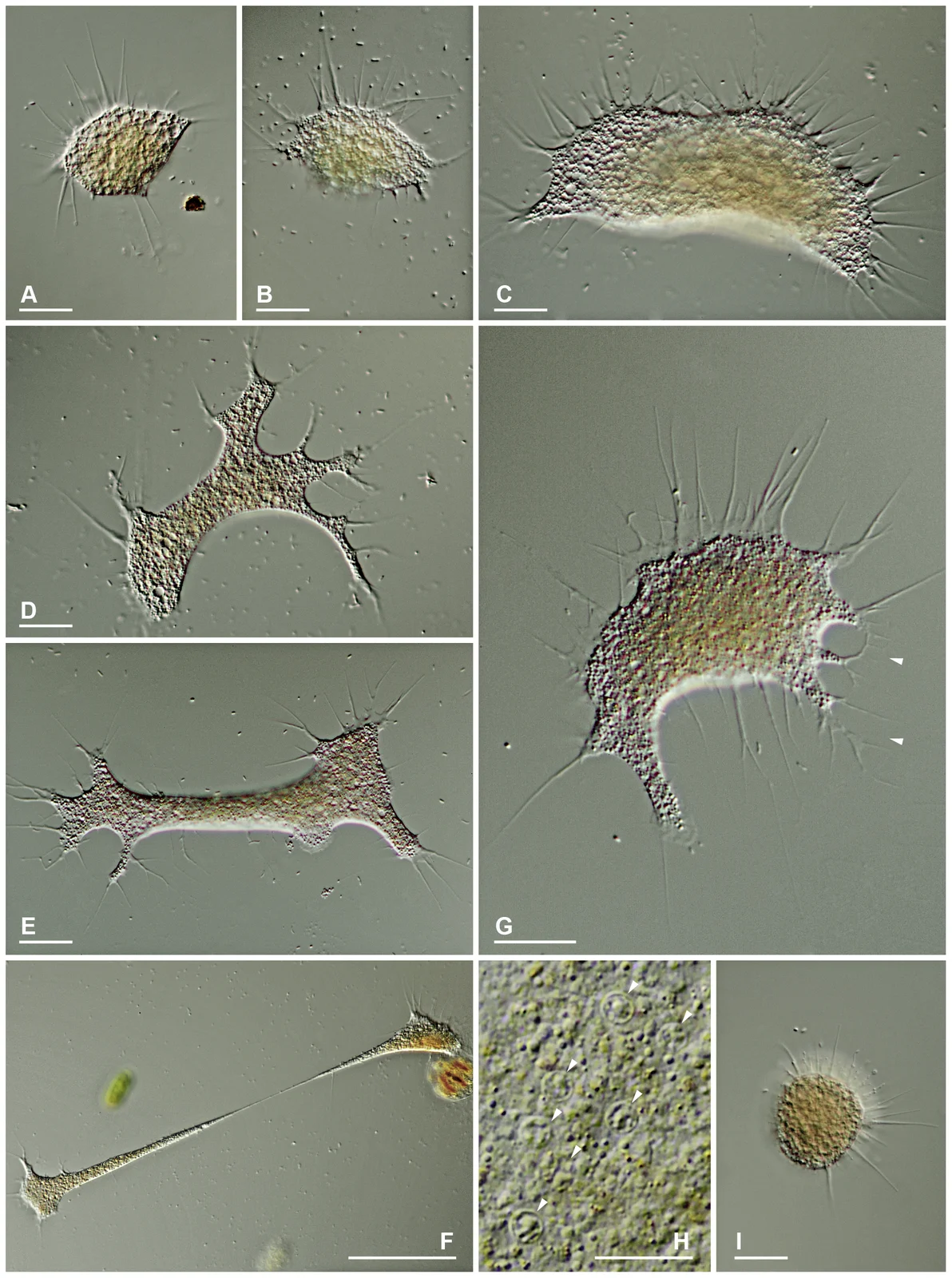
Differential-interference-contrast micrographs of Leptophrys vorax feeding cells in several shapes, Hess, Sausen and Melkonian 2012 figure4
These differential-interference-contrast micrographs show the changing outlines of Leptophrys vorax feeding cells: compact, fan-shaped, branched, elongated and floating forms. Panel H shows nuclei in a dying individual. Scale bars differ between panels; the microscopy appearance is not a claim that each cell has a fixed shape or natural color.
Overview
Leptophrys vorax is a freshwater predatory amoeba whose appearance changes markedly with its movement and food supply. The four strains examined in a 2012 study generally spread across surfaces, sometimes forming broad fans, branched bodies or long stretched strands. Their clear, slender pseudopodia emerge along the margins, occasionally from thin fringes of cytoplasm. Small moving individuals can be relatively compact, while extended cells or fused structures can exceed a millimetre. A single outline is therefore a poor basis for identifying this organism.
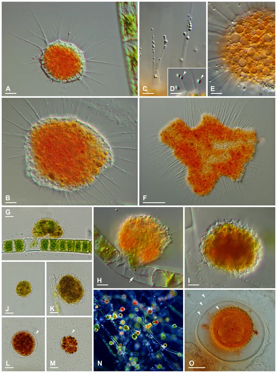
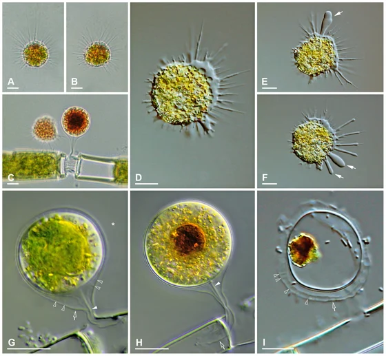
Unlike the studied Vampyrella species, which open algal walls and consume the contents, Leptophrys engulfs whole food items. Tested organisms include several desmids and yeast cells; natural observations before isolation also include diatoms and pieces of filamentous algae. Several prey cells may accumulate inside an advancing amoeba. Changing prey colors suggest that some digestion begins before the organism enters its stationary digestive-cyst phase. These observations support a broader tested feeding range, without establishing that it will consume every microorganism or animal encountered.
The digestive cyst closely reflects its contents. Small cysts can be rounded or elliptical, while those containing large algae may become elongated, lobed or dumbbell-shaped. A cyst containing a curved Closterium cell can appear banana-shaped, and another containing a slender diatom can be correspondingly narrow. Only one closely fitting envelope is observed. As digestion progresses, green inclusions become brown; remnants remain after the new mobile cells emerge. Internal division occurs, but daughter cells can fuse again during emergence, complicating the boundary between independent cells and plasmodia.
When food becomes scarce, cells may become rounded and detach into the water with radiating pseudopodia, superficially resembling Vampyrella. Large branched or connected plasmodia also occur in depleted cultures. The authors interpret some changes as possible strategies for migration or searching a wider area, but do not directly establish their ecological advantage. Multiple nuclei are difficult to distinguish in healthy living cells; visible nuclei in dying compressed specimens are not evidence that the organism has a fixed nucleus count throughout its life.
The study links this morphology with small-subunit ribosomal DNA. Four cultured strains form a distinct Leptophrys clade, separated from the sampled Vampyrella lineages. Within that clade, two sequence groupings reveal genetic divergence among strains that cannot be reliably separated by the observed morphology. The researchers refrain from naming additional species because food spectrum, ultrastructure and plasticity require further study. Their use of L. vorax reconciles older conflicting names and observations, rather than treating all similarly spreading amoebae as one confirmed species.
Origins & earliest records
The 2012 study compares cultures LV.01–LV.04 with historical descriptions and ribosomal sequences, retaining Leptophrys vorax while identifying unresolved genetic subdivisions.
Evidence & interpretation
Single-cell-derived cultures, microscopy, tested prey and ribosomal phylogeny support the account. Morphologically indistinguishable sequence groups are not automatically described as new species.
Selected bibliography
Documented works and useful reading. This is not a list of every appearance.
- 01
Shedding Light on Vampires: The Phylogeny of Vampyrellid Amoebae Revisited
PLOS ONE 7(2):e31165; complete relevant morphology, feeding, phylogeny, taxonomic discussion and culture/sequencing Methods. Original adaptation of the article’s Creative Commons Attribution license; source and authors credited.
Family, evolution & connections
Selected named vampyrellids in Hess, Sausen and Melkonian’s 2012 ribosomal tree
Six named terminals retained from Figure 1’s 81-sequence analysis. Environmental sequences, additional culture strains, other endomyxans and the filosan outgroup are pruned. The displayed split does not assert that these two sampled groups exclude intervening environmental lineages.
Scroll sideways to see all branches. With a keyboard, focus the tree and use the arrow keys.
- Selected named vampyrellid sequences
- Selected Leptophryidae
- Selected soil-derived sequences
- Platyreta germanica, AY941201
- Arachnula / Theratromyxa pair
- “Arachnula impatiens”, EU567294
- Theratromyxa weberi, ATCC50200; GQ377666
- Selected Vampyrella sequences
SSU rDNA: 1,640 aligned positions; RAxML maximum likelihood under GTR+I+Γ with 20 randomized starts. Neighbour-joining, parsimony and Bayesian analyses provide comparisons; each bootstrap analysis used 1,000 replicates. A single ribosomal locus and incomplete sampling limit this historical hypothesis. Leptophrys morphologically indistinguishable cultures include two sequence lineages; LV.03 represents only one. Support values are omitted rather than transferred onto newly collapsed nodes. Branch lengths, ages and direct ancestors are not inferred. The quoted Arachnula identification and accession remain those of the source.
Taxonomic classification
A hierarchy of classified groups, not a chain of direct ancestors.
- GenusLeptophrys
- SpeciesLeptophrys vorax
Budding yeast
Hess, Sausen and Melkonian list Saccharomyces cerevisiae among confirmed food organisms of their Leptophrys cultures, alongside several algae. These amoebae engulf whole prey cells rather than only extracting algal contents through a puncture. The observations establish a broader tested food range; they do not quantify yeast’s importance in wild diets or demonstrate identical preferences across the morphologically similar sequence lineages.
References
Sources supporting this profile. Linked pages have their own scope and editorial standards.
- ResearchHess et al. 2012 primary culture and phylogenetic studyResearch access: 2026-10-05
Image credits
Scientific organism microscopy photographDifferential-interference-contrast micrographs of Leptophrys vorax feeding cells in several shapes, Hess, Sausen and Melkonian 2012 figure4
These differential-interference-contrast micrographs show the changing outlines of Leptophrys vorax feeding cells: compact, fan-shaped, branched, elongated and floating forms. Panel H shows nuclei in a dying individual. Scale bars differ between panels; the microscopy appearance is not a claim that each cell has a fixed shape or natural color.