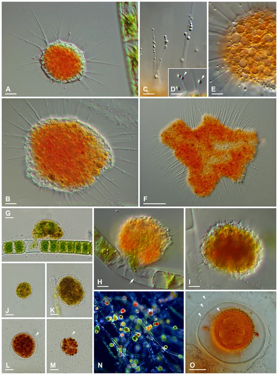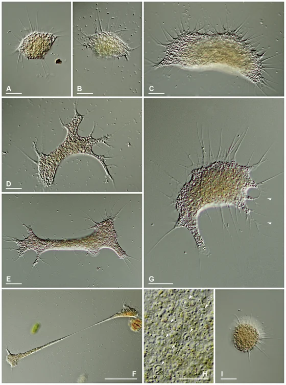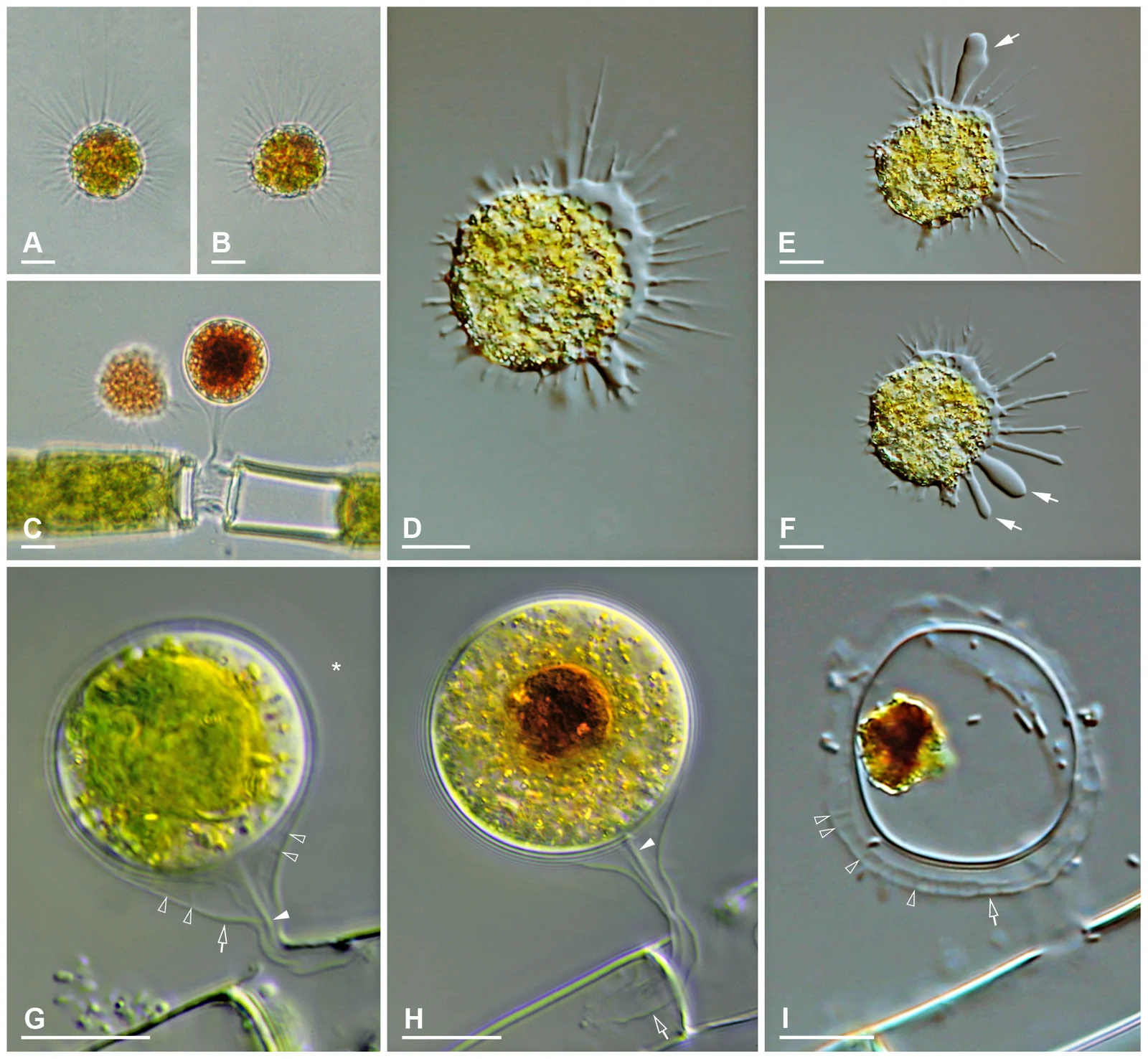
Microscopy composite of Vampyrella pendula feeding cells and digestive cyst stages, Hess, Sausen and Melkonian 2012 figure 3
The panels show mobile feeding cells and digestive cysts, including cells attached to an Oedogonium algal filament. Brightfield and differential-interference-contrast microscopy reveal different structures. Arrows, panel letters and 10-micrometre scale bars belong to the original scientific figure.
Overview
Vampyrella pendula is a freshwater amoeba that feeds on Oedogonium algae. Its compact feeding cells carry long, clear, thread-like pseudopodia around an orange central body. In the strains examined in 2012, young cells are about 20–30 micrometres across and pseudopodia can extend to about 50 micrometres. The projections can branch or occur in tufts. Unlike the compared Vampyrella lateritia cells, they do not show moving refractive granules along the pseudopodia. Cells can float freely, stalk over surfaces or make paddling movements.
After feeding, the organism forms a digestive cyst attached to its food alga. This stage gives the species a particularly recognizable appearance: the outer envelope extends into a hollow stalk anchored within an emptied algal cell. The cyst itself is approximately spherical, about 25 micrometres in the examined material. Three envelopes can be distinguished, although some are delicate and difficult to see. A clear strand runs from the innermost envelope through the stalk toward its attachment, an observed structural feature rather than a demonstrated transport mechanism.
Digestion alters the color of the cyst contents. Early stages contain green material surrounded by paler cytoplasm; mature stages become orange-red and contain a brown food remnant. After emergence, that remnant stays inside the abandoned envelope. Feeding generally resembles the localized algal-wall opening described for V. lateritia, but the investigators do not observe V. pendula consuming several algal cells successively. Resting cysts appear, although their structure is not examined in comparable detail. Cell fusion into plasmodia is not observed in this study.
Two strains, VP.01 and VP.02, are identified by comparison with earlier descriptions and supported as a closely related sequence pair. Feeding succeeds with the tested Oedogonium stellatum and O. cardiacum cultures. The strains do not consume the offered zygnematalean algae eaten by V. lateritia. Conversely, the latter fails to eat the tested Oedogonium. These differences suggest separation of feeding niches between related amoebae, but the trials establish a tested preference rather than a complete nutritional range.
The broader phylogenetic study uses ribosomal DNA and cultured morphology to distinguish Vampyrella from more spreading, often omnivorous Leptophrys forms. Their shared digestive-cyst life stages do not make them slime moulds, and superficially similar thread-bearing amoebae need not be close relatives. The paper also emphasizes that food supply can change cellular shape. Reliable identification therefore draws on multiple individuals and life stages under suitable conditions, instead of one photograph or a single cell’s outline.
Origins & earliest records
The 2012 study compares single-cell-derived strains VP.01 and VP.02 with historical descriptions and small-subunit ribosomal DNA. The research cultures are not newly designated original types.
Evidence & interpretation
Microscopy documents feeding cells and stalked digestive cysts; culture trials and ribosomal sequences distinguish the sampled organism from V. lateritia. Resting cysts were not examined in detail.
Selected bibliography
Documented works and useful reading. This is not a list of every appearance.
- 01
Shedding Light on Vampires: The Phylogeny of Vampyrellid Amoebae Revisited
PLOS ONE 7(2):e31165; complete relevant morphology, feeding, phylogeny, taxonomic discussion and culture/sequencing Methods. Original adaptation of the article’s Creative Commons Attribution license; source and authors credited.
Family, evolution & connections
Selected named vampyrellids in Hess, Sausen and Melkonian’s 2012 ribosomal tree
Six named terminals retained from Figure 1’s 81-sequence analysis. Environmental sequences, additional culture strains, other endomyxans and the filosan outgroup are pruned. The displayed split does not assert that these two sampled groups exclude intervening environmental lineages.
Scroll sideways to see all branches. With a keyboard, focus the tree and use the arrow keys.
- Selected named vampyrellid sequences
- Selected Leptophryidae
- Selected soil-derived sequences
- Platyreta germanica, AY941201
- Arachnula / Theratromyxa pair
- “Arachnula impatiens”, EU567294
- Theratromyxa weberi, ATCC50200; GQ377666
- Selected Vampyrella sequences
SSU rDNA: 1,640 aligned positions; RAxML maximum likelihood under GTR+I+Γ with 20 randomized starts. Neighbour-joining, parsimony and Bayesian analyses provide comparisons; each bootstrap analysis used 1,000 replicates. A single ribosomal locus and incomplete sampling limit this historical hypothesis. Leptophrys morphologically indistinguishable cultures include two sequence lineages; LV.03 represents only one. Support values are omitted rather than transferred onto newly collapsed nodes. Branch lengths, ages and direct ancestors are not inferred. The quoted Arachnula identification and accession remain those of the source.
Taxonomic classification
A hierarchy of classified groups, not a chain of direct ancestors.
- GenusVampyrella
- SpeciesVampyrella pendula
References
Sources supporting this profile. Linked pages have their own scope and editorial standards.
- ResearchHess et al. 2012 primary culture and phylogenetic studyResearch access: 2026-10-05
Image credits
Scientific organism microscopy photographMicroscopy composite of Vampyrella pendula feeding cells and digestive cyst stages, Hess, Sausen and Melkonian 2012 figure 3
The panels show mobile feeding cells and digestive cysts, including cells attached to an Oedogonium algal filament. Brightfield and differential-interference-contrast microscopy reveal different structures. Arrows, panel letters and 10-micrometre scale bars belong to the original scientific figure.