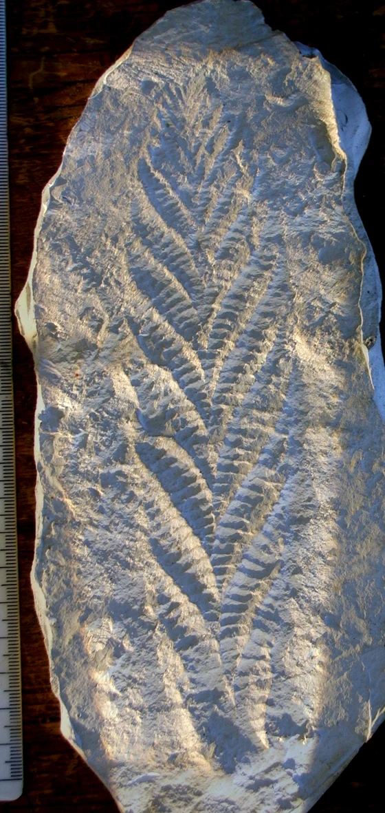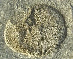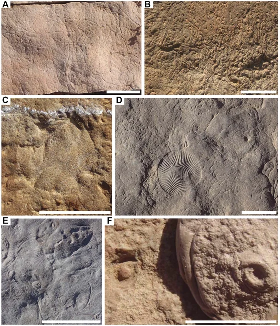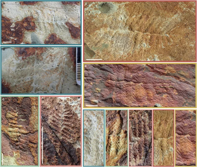
Fractofusus andersoni fossil specimens and enlarged branching details in Dunn, Donoghue and Liu’s figure 4
Field photographs show the central pair of branches in Fractofusus andersoni, with enlarged details below. Panels A and B show the same specimen; the figure’s scale bars represent one centimetre. These are fossil impressions in rock, rather than a reconstruction of the organism’s living appearance. The published figure is reproduced unchanged.
Overview
Fractofusus andersoni had two tapering ends and paired rows of branching units along a narrow central axis. Its largest branches lay near the middle, becoming shorter toward the tips. Each first-order branch contained one small frond, whereas the other recognized species, F. misrai, bundled multiple fronds into comparable branches. These repeated structures give the fossil a superficially leafy appearance, but that resemblance does not classify it as a plant. Dunn, Donoghue and Liu interpret rangeomorphs as early members of the animal lineage leading toward eumetazoans; their exact internal relationships and feeding functions remain debated.
Their 2025 study examined more than 100 fossils on the Brasier Surface at Mistaken Point, using field observations and detailed casts. Eighteen especially informative specimens, measuring 7.8–69.7 mm, supplied the quantitative growth dataset. Larger individuals generally had more first-order branches, while existing branches also enlarged. Both tips contributed new branches. The authors infer a shift from predominantly adding branches toward increasing the size of existing ones, producing a more diamond-shaped outline during later growth. The smallest observed fossil already possessed 18 first-order branches, so the proposed earliest two-branch stage remains a reconstruction rather than a directly preserved juvenile.
Several fossils terminate abruptly, while some retain an extension of the central axis beyond their branching body. The authors interpret the continuous axis as a runner-like structure, or stolon. This supports comparison with connected or clonal rangeomorphs, but does not settle whether the whole body should be understood as an integrated colony. Nor can every truncated fossil be explained confidently: physical damage, budding and fragmentation remain competing possibilities. The examined broken specimens do not show regrowth from their damaged tips.
Comparison with Charnia, Bradgatia and other rangeomorphs places these observations in a broader question about how early animal bodies developed. Shared branching principles could explain differences in the length and arrangement of their main axes, without making any named genus the direct ancestor of another. The study argues that these bodies required substantial developmental regulation before familiar animal organs appeared. This is an inference from organized fossil growth, not the recovery of Ediacaran genes. Because the analysis concerns one surface and one species, the authors explicitly avoid pooling different fossil beds or treating their results as measurements of every Fractofusus population.
Origins & earliest records
Gehling and Narbonne named Fractofusus andersoni in 2007. The 2025 developmental study sampled Briscal Formation bed BR5 within the Mistaken Point Ecological Reserve. Its quoted age, 567.63 ± 0.66 million years, dates the studied surface rather than the entire stratigraphic range of the genus.
Evidence & interpretation
Dunn, Donoghue and Liu (2025), Results, Discussion and Methods, document the central axis, branch organization and growth measurements. Studied casts and molds are held by the Sedgwick Museum, CAMSM X.50400.1–X.50400.17; these accessioned research materials are not identified here as the species holotype. Tectonic distortion was corrected before analysis. Higher branching orders were excluded where preservation prevented comparison, and the fossil surface may represent a time-averaged community.
Selected bibliography
Documented works and useful reading. This is not a list of every appearance.
- 01
Morphogenesis of Fractofusus andersoni and the nature of early animal development
Nature Communications 16, article 3439; complete Results, Discussion and Methods. Published 11 April 2025. Original summary adapted from an article licensed CC BY 4.0; https://creativecommons.org/licenses/by/4.0/.
- 02
Spindle-shaped Ediacara fossils from the Mistaken Point assemblage, Avalon Zone, Newfoundland
Canadian Journal of Earth Sciences44,367–387; DOI10.1139/E07-003
Family, evolution & connections
Taxonomic classification
A hierarchy of classified groups, not a chain of direct ancestors.
- GenusFractofusus
- SpeciesFractofusus andersoni
Located primary passages
Specific passages supporting details in this entry, grouped by their published witness.
Primary source witness
Canadian Journal of Earth Sciences44,367–387; DOI10.1139/E07-003
- The original species diagnosis distinguishes an ovate spindle with at most thirteen modules per vane from the more elongated F. misrai. Holotype ROM38636 comes from Bristy Cove’s Briscal Formation; the name honours Michael Anderson. The authors’ growth interpretation relies on module counts and reconstructed fossil shape, with unchanged ventral/dorsal structure assumed by comparison rather than directly demonstrated in folded F. andersoni.
Species diagnosis/material379; continuation and comparison380–381; Fig.12
No whole corpus or uninspected supplementary materials claimed; research hypotheses distinguished from observation. No quotation or image reuse.
References
Sources supporting this profile. Linked pages have their own scope and editorial standards.
- ResearchDunn, Donoghue and Liu (2025), developmental analysis of Fractofusus andersoniResearch access: 2026-10-05
Image credits
Scientific fossil specimen photographFractofusus andersoni fossil specimens and enlarged branching details in Dunn, Donoghue and Liu’s figure 4
Field photographs show the central pair of branches in Fractofusus andersoni, with enlarged details below. Panels A and B show the same specimen; the figure’s scale bars represent one centimetre. These are fossil impressions in rock, rather than a reconstruction of the organism’s living appearance. The published figure is reproduced unchanged.