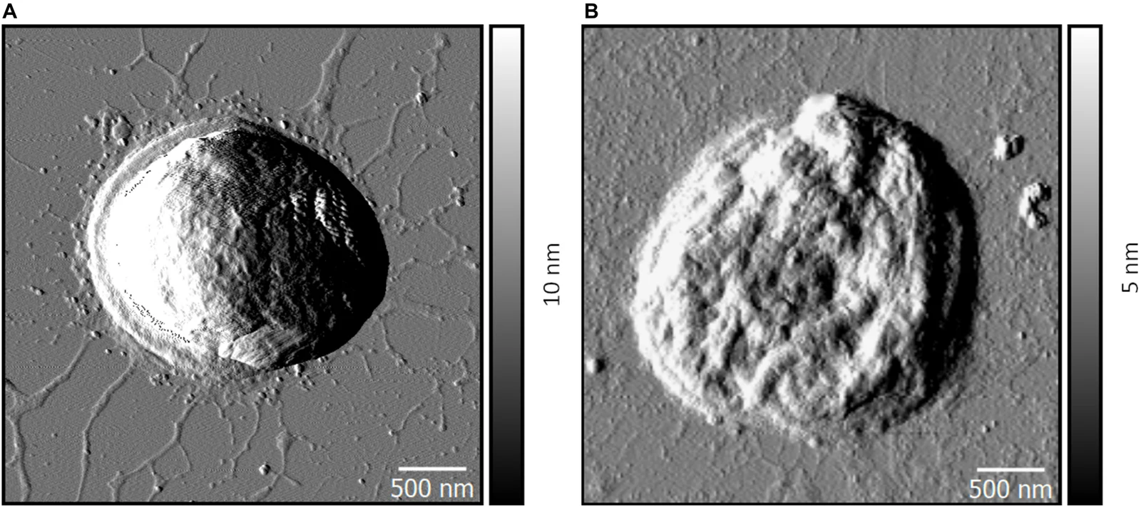
Atomic-force microscopy amplitude images of wild-type and ΔpilD* Synechocystis sp. PCC 6803 cells in Thirumurthy and colleagues’ 2020 figure 3
Panel A shows a wild-type Synechocystis sp. PCC 6803 cell with surrounding filaments; panel B shows the experimental ΔpilD* strain. These are atomic-force microscopy amplitude images, with 500-nanometre scale bars. Their greyscale contrast records the microscopy signal rather than natural cell colour. The panels compare particular experimental strains.
Overview
Synechocystis sp. PCC 6803 is a laboratory cyanobacterial strain used to investigate photosynthesis. Its light-harvesting apparatus includes phycobilisomes: assemblies of proteins carrying colored molecules called bilins. These antennae absorb light and pass excitation energy toward photosynthetic reaction centers. A smaller antenna type, CpcL-phycobilisome, consists of a rod without the allophycocyanin core found in larger complexes. Lvqin Zheng and colleagues combined cryogenic electron microscopy with ultrafast spectroscopy in 2023 to examine how this relatively compact apparatus could deliver absorbed energy to photosystem I.
The preparation came from an engineered PCC 6803 strain lacking apcAB, which helped isolate the antenna under study. Cultures were grown under defined light and temperature conditions, and purified complexes were stabilized before freezing for microscopy. The dominant reconstructed particle contained three stacked protein hexamers; other rod lengths occurred in the preparation. A density map at approximately 2.6 angstrom resolution supported models of protein chains and their attached pigments. The rod was about 160 angstroms long and 100 angstroms across, showing the scale of a molecular assembly rather than the dimensions of a whole bacterium.
Four linker elements formed a supporting arrangement through the rod. At its distal end, the team resolved the CpcD domain of ferredoxin–NADP oxidoreductase. The enzyme's other two domains were not resolved, which the authors associated with their flexible connection. At the membrane-facing end, the transmembrane region of CpcL was likewise unresolved. These distinctions matter: the study combined identifiable structural features with proposed positioning in the photosynthetic membrane, rather than directly imaging every part of an intact living-cell supercomplex.
The protein environment altered the shapes of particular bilins. One pigment near the membrane-facing end was especially flattened and was assigned a role as the red-shifted terminal energy acceptor. Time-resolved absorption and fluorescence measurements supported rapid energy movement among different components, followed by a longer-lived terminal component. Assignments of some individual pigments were tentative, and the fluorescence components depended on the analytical model used to separate overlapping signals. The structural and spectroscopic evidence together provided a mechanism for directed light-energy transfer. It did not establish that every cyanobacterial species uses an identical antenna arrangement or directly reconstruct the original evolution of photosynthesis.
Origins & earliest records
PCC 6803 laboratory strain; the focal structural study used an engineered apcAB deletion derivative.
Evidence & interpretation
Purified antenna complexes were investigated by cryo-EM and time-resolved spectroscopy; pigment assignments and flexible unresolved regions retain their qualifications.
Selected bibliography
Documented works and useful reading. This is not a list of every appearance.
- 01
Cryo-EM and femtosecond spectroscopic studies provide mechanistic insight into the energy transfer in CpcL-phycobilisomes
Nature Communications 14:3961; DOI 10.1038/s41467-023-39689-7. Complete main report and Methods; CC BY 4.0.
Family, evolution & connections
Taxonomic classification
A hierarchy of classified groups, not a chain of direct ancestors.
- kingdomBacteria
- phylumCyanobacteria
- genusSynechocystis
- strainPCC 6803
References
Sources supporting this profile. Linked pages have their own scope and editorial standards.
- ResearchZheng and colleagues 2023, primary structural and spectroscopic report, CC BY 4.0Research access: 2026-10-05
Image credits
Scientific organism atomic-force microscopy figureAtomic-force microscopy amplitude images of wild-type and ΔpilD* Synechocystis sp. PCC 6803 cells in Thirumurthy and colleagues’ 2020 figure 3
Panel A shows a wild-type Synechocystis sp. PCC 6803 cell with surrounding filaments; panel B shows the experimental ΔpilD* strain. These are atomic-force microscopy amplitude images, with 500-nanometre scale bars. Their greyscale contrast records the microscopy signal rather than natural cell colour. The panels compare particular experimental strains.