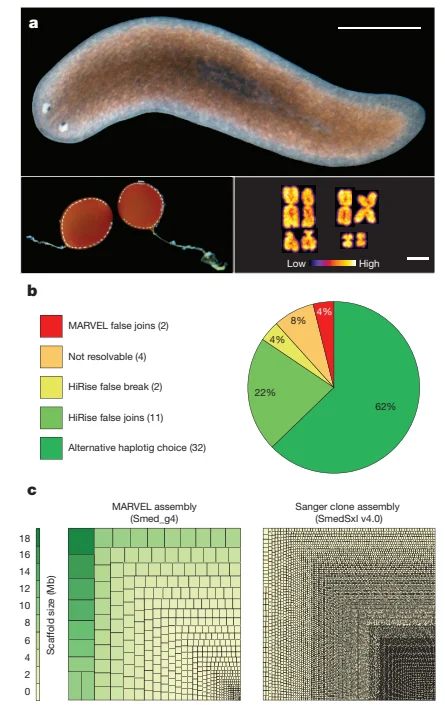
Sequenced sexual-strain Schmidtea mediterranea individual, egg cocoons and karyotype in panel a, with genome-assembly charts below, Grohme and colleagues’ 2018 figure 1
Panel A shows an individual of the sequenced sexual Schmidtea mediterranea strain, with egg cocoons and a karyotype below. Scale bars represent two millimetres for the animal and 2.5 micrometres for the karyotype. The chromosome colours encode the image rather than natural pigmentation. Panels B and C show assembly quality and scaffold comparisons, not additional animal specimens. The complete figure was extracted from the published PDF.
Overview
Schmidtea mediterranea is a free-living planarian flatworm whose capacity to replace lost tissues makes it an important experimental animal. Adult stem cells called neoblasts support regeneration and ordinary tissue turnover. The species belongs to the same broad flatworm radiation as parasitic flukes and tapeworms, but its free-living biology provides a different comparative setting. Its value in the selected study lies in testing cellular mechanisms in an animal that retains complex regenerative functions despite substantial genomic differences from more familiar laboratory species.
Grohme and colleagues assembled a reference genome from a sexual laboratory strain that had undergone more than seventeen generations of sibling mating. They combined long-read sequencing, a new assembler designed for difficult sequence and proximity-ligation scaffolding. The resulting assembly contained 782.1 million bases in 481 scaffolds. More than 99 percent of the strain’s transcripts mapped back to it, supporting extensive coverage rather than proving that every sequence was correct. Comparison of assembly and scaffolding detected structural conflicts, while small duplications and uncertain regions near gaps remained. This is a particular experimental reference, not a complete inventory of variation throughout the species.
Several features help explain why earlier assemblies were fragmented. The genome has abundant low-complexity, AT-rich sequence and substantial variation that persisted despite inbreeding. Repetitive elements account for much of the assembly. Three unusually long retroelement families, named Burro, exceeded thirty thousand bases and contributed disproportionately to difficult scaffold boundaries. Expression of retroelement sequence does not itself establish that every copy is active. Comparisons with other animals also identified numerous apparently missing conserved genes, including homologues considered essential in particular human or mouse experiments. Such comparisons depend on available assemblies and on the ability to recognize divergent homologues; they do not establish that a cellular function is absent whenever its familiar gene is not found.
The spindle assembly checkpoint illustrates that distinction. Recognizable MAD1 and MAD2 homologues were not identified in the planarian data, yet the drug nocodazole still caused cells to arrest during mitosis. Gene-silencing experiments targeting components of the ROD–ZW10–ZWILCH complex prevented this drug-induced arrest without reducing basal stem-cell numbers or proliferation in those tests. The authors proposed that checkpoint control could use a different arrangement of components, or homologues whose sequence had diverged beyond recognition. The experiment therefore links a molecular perturbation to an observed response, while leaving the complete mechanism unresolved. It does not show that every neoblast behaves identically or that genome comparison alone explains regeneration.
Origins & earliest records
The selected reference represents an inbred sexual laboratory strain. Its 2018 publication is a research date, not an evolutionary origin date.
Evidence & interpretation
Long-read assembly, transcript mapping, comparative gene searches and RNA-interference perturbations provide distinct lines of evidence. Apparent gene absence and proposed alternative checkpoint mechanisms remain qualified interpretations.
Selected bibliography
Documented works and useful reading. This is not a list of every appearance.
- 01
The genome of Schmidtea mediterranea and the evolution of core cellular mechanisms
Nature 554: 56–61; online 24 January, issue 1 February. Complete main report and selected main/extended caption texts read; DOI 10.1038/nature25473. Published PDF explicitly CC BY 4.0 despite generic copyright footers.
Family, evolution & connections
Selected flatworm protein-sequence branching (Grohme et al., 2018)
All eight flatworm terminals from Figure 3A, retaining sexual and asexual Schmidtea mediterranea samples separately, with the oyster as one selected lophotrochozoan outgroup. Other animals are omitted.
Scroll sideways to see all branches. With a keyboard, focus the tree and use the arrow keys.
- Selected flatworms and oyster
- Sampled flatworms
- Remaining sampled flatworm branches
- Schmidtea mediterranea samples
- Selected parasitic flatworms
- Schistosoma mansoni
- Selected cestodes
- Hymenolepis microstoma
- Remaining sampled cestodes
- Taenia solium
- Echinococcus pair
- Echinococcus granulosus
- Echinococcus multilocularis
- Macrostomum lignano
- Oyster — C. gigas (source label), selected outgroup
Supplementary S14.1 describes OrthoMCL orthologue selection across 22 species, PRANK alignment, trimAl filtering and concatenation, followed by RAxML 8.2.9 with PROTCATLG. Non-flatworm relationships were constrained to a guide tree. Figure 3A calls the dataset 51 single-copy genes; S14.1 instead describes 52 conserved proteins. That discrepancy is retained rather than reconciled. This reproduces the displayed flatworm branching, not a new inference. No node support values are printed in Figure 3A, so none are supplied here. Its scale is substitutions per site, not time; branch lengths are omitted. Sexual/asexual samples belong to one species. Gene-loss comparisons in panel 3C do not prove that parasitism caused each loss. Adapted from Grohme et al. (2018), CC BY 4.0: nine terminals retained, omitted branches suppressed, layout redrawn and lengths omitted.
References
Sources supporting this profile. Linked pages have their own scope and editorial standards.
- ResearchPublished genome and cellular-mechanism studyResearch access: 2026-10-05
Image credits
Scientific animal photograph, karyotype and genome-assembly figureSequenced sexual-strain Schmidtea mediterranea individual, egg cocoons and karyotype in panel a, with genome-assembly charts below, Grohme and colleagues’ 2018 figure 1
Panel A shows an individual of the sequenced sexual Schmidtea mediterranea strain, with egg cocoons and a karyotype below. Scale bars represent two millimetres for the animal and 2.5 micrometres for the karyotype. The chromosome colours encode the image rather than natural pigmentation. Panels B and C show assembly quality and scaffold comparisons, not additional animal specimens. The complete figure was extracted from the published PDF.