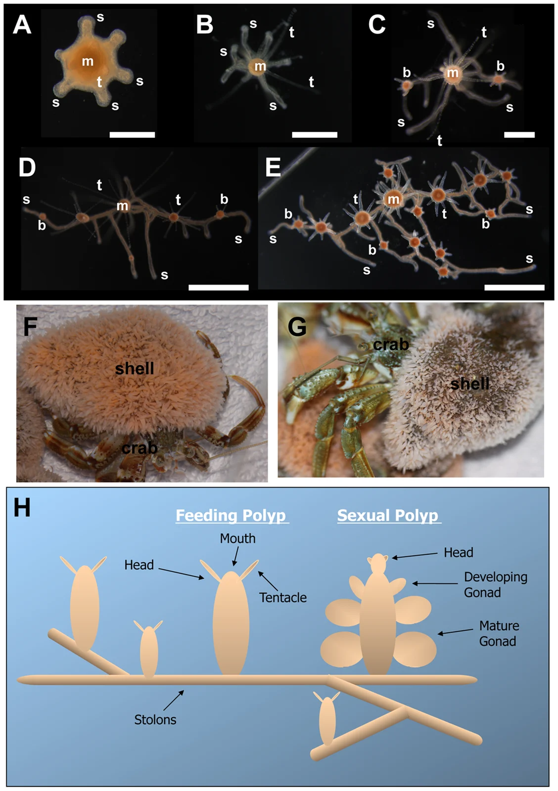
Hydractinia echinata colony micrographs, colonies covering hermit-crab shells, and a labelled feeding and sexual polyp diagram.
Whole published Figure 1: A–E show early colony growth; F–G show mature colonies covering hermit-crab shells; H is a schematic, rather than a photograph.
Overview
Hydractinia echinata forms colonies of small marine polyps connected by gastrovascular tubes called stolons. The selected study describes it as common in the European North Atlantic and useful for regeneration research because its translucent, attached polyps can be cultured and followed microscopically. Colonies can grow by stolon extension and by budding additional polyps, alongside sexual reproduction. A polyp and the stolon network are therefore distinct structures within a continuing colony, rather than interchangeable names for the same body part.
Bradshaw, Thompson and Frank examined what happened after removing heads and separating polyps from their colonies. Their laboratory animals were maintained in artificial seawater at 18°C and fed brine shrimp and ground fish. Head wounds closed within hours, primarily through epithelial stretching. A new mound of tissue, or blastema, appeared at the oral end, followed by a mouth and tentacles. Most tested animals regained feeding ability, although the timing varied with age, genetic background and health. Formation of a head over days did not coincide with direct restoration of the removed basal stolons.
Two complementary approaches traced the cellular source of the head blastema. A pulse of labelled DNA precursor marked dividing cells in the lower polyp before decapitation; labelled cells later appeared in the developing head. In separately engineered animals, a fluorescent reporter associated with Piwi1 expression allowed individual cells to be followed as they migrated and sometimes divided. These results supported movement of interstitial stem cells from a more distant region. They did not exclude a contribution from existing epithelial cells. Irradiation that suppressed proliferation also prevented blastema formation and head regeneration, supporting a requirement for proliferating cells in this experimental setting.
The basal response was strikingly different. Isolated polyps initially retained their form and could feed, but over subsequent weeks some lost tentacles and the rest of the head, elongated and developed branches. The transformed tissue secreted chitin and budded new polyps, eventually forming sexually mature colonies. This was interpreted as conversion of a polyp into stolon tissue, followed by rebuilding polyps, rather than immediate growth of a stolon from the wound. Loss of localized Wnt3 expression accompanied loss of head identity. The authors proposed a connection between tissue polarity and the different regenerative routes, while leaving the precise causal mechanism unresolved. Their observations establish substantial tissue-level developmental flexibility; they do not determine whether every participating stem cell is individually pluripotent.
Origins & earliest records
A living colonial hydrozoan represented by a polymorphic laboratory population in the selected experiment; research publication does not date its evolutionary origin.
Evidence & interpretation
Labelled-cell tracing, transgenic reporter observations, proliferation perturbation and long-term morphology provide complementary evidence. Epithelial contributions, single-cell potency and the causal role of polarity remain incompletely resolved.
Selected bibliography
Documented works and useful reading. This is not a list of every appearance.
- 01
Distinct mechanisms underlie oral vs aboral regeneration in the cnidarian Hydractinia echinata
eLife 4: e05506. Complete Introduction, Discussion and Materials and Methods; selected head, proliferation requirement, migration and stolon Results. DOI 10.7554/eLife.05506. CC BY 4.0.
Family & connections
A source-supported family tree or inspiration record has not yet been compiled for this profile. Related reading below indicates a connection in subject matter, not a proven lineage or creative influence.
Look for a taxonomic record and classified relativesLocated primary passages
Specific passages supporting details in this entry, grouped by their published witness.
Primary source witness
2015 PMC text
- This study of Hydractinia echinata distinguishes head regeneration from rebuilding a colony after stolon removal. Labelled cells and transgenic reporters support migration of interstitial stem cells from the lower body into a proliferating head blastema. Irradiation and proliferation inhibition impaired regeneration. Knocking down selected stem-cell or nematogenesis genes impaired head restoration without preventing blastema formation, separating those outcomes. In contrast, isolated polyps did not directly regenerate stolons at their aboral cut. They lost head structures, transformed into stolonal tissue and subsequently budded feeding and sexual polyps. The experiments therefore support tissue-specific regenerative mechanisms within one species.
The authors did not resolve pluripotent individual cells versus multiple lineage-restricted progenitors; epithelial contributions were not excluded. No independently watched microscopy videos, figure-pixel inspection, supplementary datasets, rerun or human medical application is claimed.
References
Sources supporting this profile. Linked pages have their own scope and editorial standards.
- ResearchOral and aboral regeneration experimentsResearch access: 2026-10-05
Image credits
Organism microscopy, shell-colony photographs and explanatory anatomy diagramHydractinia echinata colony micrographs, colonies covering hermit-crab shells, and a labelled feeding and sexual polyp diagram.
Whole published Figure 1: A–E show early colony growth; F–G show mature colonies covering hermit-crab shells; H is a schematic, rather than a photograph.