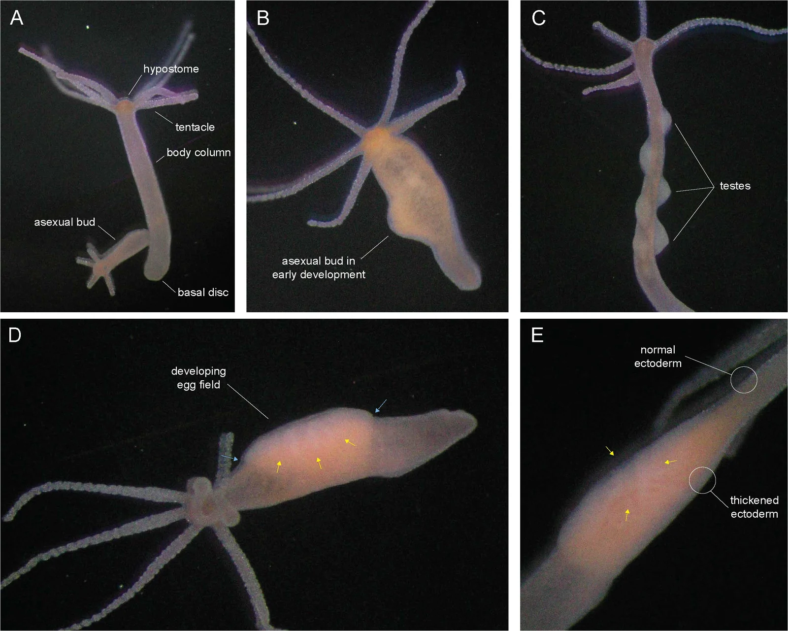
Five labelled photographs of laboratory Hydra vulgaris polyps showing asexual buds, testes and a developing egg field, Cochran and colleagues’ 2026 figure 1
These laboratory Hydra vulgaris photographs show a developed asexual bud in A, an early bud in B, testes in C, and a developing oocyte and egg field in D. Panel E compares the body column with thickened tissue around the egg field. Panels A–D were photographed at 3.2× magnification and E at 5×. The labels and coloured arrows identify anatomical features in these study animals.
Overview
Cazet and colleagues assembled a chromosome-level reference for the AEP laboratory strain of Hydra vulgaris. Long-read sequencing, linked reads and chromosome-contact data supported assembly and annotation. Comparison with strain 105 found broadly conserved chromosome organization, with an approximately five-million-base inversion on chromosome eight. These laboratory references do not survey all wild variation.
Chromatin-accessibility assays, histone-mark mapping and cross-species sequence comparisons identified candidate regulatory regions. Predicted binding sites were computationally filtered; their presence does not demonstrate every proposed regulatory interaction. The resource supports investigation of epithelial and interstitial stem-cell differentiation, while detailed experimental protocols and datasets require separate examination.
Origins & earliest records
AEP reference study published in 2023.
Evidence & interpretation
Genome and chromatin assays; regulatory function partly inferred.
Selected bibliography
Documented works and useful reading. This is not a list of every appearance.
- 01
A chromosome-scale epigenetic map of the Hydra genome reveals conserved regulators of cell state
Genome Research 33: 283–298. Selected genome/regulatory Results and complete main Methods. CC BY-NC 4.0.
Family, evolution & connections
AEP and 105 genomes
Chromosomal synteny was broadly conserved between these laboratory strains.
References
Sources supporting this profile. Linked pages have their own scope and editorial standards.
- ResearchCazet 2023Research access: 2026-10-05
Image credits
Scientific animal anatomy photograph compositeFive labelled photographs of laboratory Hydra vulgaris polyps showing asexual buds, testes and a developing egg field, Cochran and colleagues’ 2026 figure 1
These laboratory Hydra vulgaris photographs show a developed asexual bud in A, an early bud in B, testes in C, and a developing oocyte and egg field in D. Panel E compares the body column with thickened tissue around the egg field. Panels A–D were photographed at 3.2× magnification and E at 5×. The labels and coloured arrows identify anatomical features in these study animals.