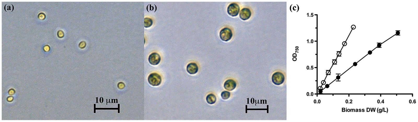
Chlorella vulgaris CCAP 211/11B cell micrographs under two culture conditions, with 10 micrometer scale bars and an optical-density versus biomass graph
Panels A and B show Chlorella vulgaris cells in unsupplemented Tris-Phosphate medium and medium containing 100 mM glucose, respectively. The 10 μm bars indicate scale. Panel C compares optical density with biomass dry weight; these cultured cells illustrate an experimental comparison rather than a single universal cell size.
Overview
Chlorella vulgaris is a freshwater green microalga investigated for its rapid growth and photosynthetic production of biomass. Cecchin and colleagues studied strain 211/11P in 2019, linking a genome assembly with measurements of cultures grown under two light intensities. This gives a concrete picture of how one cultivated strain responds to its environment, rather than an assumption that all Chlorella isolates have identical genomes or physiology. The cells were grown photoautotrophically, using light, under controlled temperature and a daily light-dark cycle.
Under high light, the cultures grew faster and accumulated more dry biomass than under low light. The average cell diameter remained similar, while individual-cell composition changed. Chlorophyll per cell decreased and the carotenoid-to-chlorophyll ratio increased. Lipid accumulation rose strongly, particularly the storage lipid triacylglycerol: its share of total lipid changed from 12% under low light to 79% under high light. Starch and protein per cell did not differ significantly. These are measured responses in the tested culture conditions, not evidence that increasing light always improves growth in every setting.
The reference genome combined long sequencing reads, more accurate short reads and optical maps of DNA. Fourteen large scaffolds contained 98.9% of the assembled nuclear sequence, with additional unplaced contigs. The authors suggested that these fourteen reconstructed molecules might correspond to chromosomes, but did not establish the chromosome count through an independent cytological observation. RNA sequencing supported annotation of 10,724 genes and 11,082 transcripts. A conserved-gene benchmark recovered 95.4% complete orthologs; this tests one aspect of completeness, rather than proving that every genomic region and gene was resolved.
The chloroplast and mitochondrial genomes were assembled separately, at 165,504 and 91,560 base pairs respectively. The mitochondrial assembly included a fragment related to a chloroplast polymerase gene, interpreted as evidence of inter-organelle transfer; its possible function remained unclear. Comparing high- and low-light cultures identified changes in transcript abundance, including reduced expression of many light-harvesting antenna genes under high light. The proposed pathways connecting carbon metabolism with lipid storage were models based on genomic and expression evidence. Their detailed enzyme roles still required experimental validation. Likewise, finding genes associated with meiosis and motility did not demonstrate mating or gamete formation in these cultures.
Origins & earliest records
The cited reference concerns a living culture-collection strain, 211/11P. Genome similarity and a comparison using 111 shared single-copy genes place it with sampled green algal relatives, including other Chlorella genomes; this does not identify a direct ancestor or date the species origin.
Evidence & interpretation
Hybrid genome assembly, RNA sequencing and controlled light-acclimation measurements support the account. Scaffold structure, conserved-gene completeness, transcript changes and biochemical measurements answer different questions. Predicted enzyme functions, chromosome correspondence and the metabolic model are explicitly interpretive.
Selected bibliography
Documented works and useful reading. This is not a list of every appearance.
- 01
Chlorella vulgaris genome assembly and annotation reveals the molecular basis for metabolic acclimation to high light conditions
The Plant Journal 100(6):1289–1305; DOI 10.1111/tpj.14508. Complete Introduction, growth/genome/organelle/expression Results, photosynthesis section, Discussion and Experimental procedures read; other pathway Results and supplements not fully read. CC BY 4.0; attributed original summary.
Family, evolution & connections
Taxonomic classification
A hierarchy of classified groups, not a chain of direct ancestors.
- genusChlorella
- speciesChlorella vulgaris
References
Sources supporting this profile. Linked pages have their own scope and editorial standards.
- ResearchChlorella vulgaris genome assembly and annotation reveals the molecular basis for metabolic acclimation to high light conditionsResearch access: 2026-10-06
Image credits
Scientific organism microscopy experiment figureChlorella vulgaris CCAP 211/11B cell micrographs under two culture conditions, with 10 micrometer scale bars and an optical-density versus biomass graph
Panels A and B show Chlorella vulgaris cells in unsupplemented Tris-Phosphate medium and medium containing 100 mM glucose, respectively. The 10 μm bars indicate scale. Panel C compares optical density with biomass dry weight; these cultured cells illustrate an experimental comparison rather than a single universal cell size.