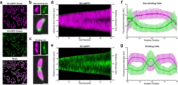
Fluorescently labelled engineered Caulobacter crescentus cells and S-layer signal profiles, Herdman and coauthors2024figure1
The micrographs show engineered Caulobacter crescentus cells labelled to compare existing and newly incorporated surface-layer material. Magenta and green encode fluorescent markers, not natural bacterial colors. The authors filtered the images to reduce noise; the surrounding plots quantify signal location in dividing and non-dividing cells.
Overview
Caulobacter crescentus is a bacterial model for cell growth, polarity and division. Its life cycle includes a non-dividing swarmer stage and a division-competent stalked stage. A protein surface layer, or S-layer, covers the cell and its stalk outside the outer membrane. This layer is formed from repeating RsaA proteins arranged in a hexagonal lattice and anchored through lipopolysaccharide. A 2024 investigation used modified laboratory cells to follow where newly produced material enters this covering.
Researchers labeled existing surface material with one fluorescent marker and subsequently added a differently colored marker during growth. The approach distinguished old and new regions rather than treating all surface fluorescence as equivalent. Short, non-dividing cells predominantly added material at their poles, while dividing cells showed conspicuous addition around the middle. Previously incorporated lattice remained comparatively stationary relative to the cell. The results connect surface renewal with changing cell geometry and growth stage, rather than describing a covering that is assembled once and then remains unchanged.
Drug perturbations helped separate related processes. Disrupting cell division with cephalexin or mitomycin C did not abolish the distinct localization of newly added surface material. In contrast, treatment affecting the cytoskeletal protein MreB or the lipopolysaccharide component disrupted that pattern. MreB inhibition also changes several cellular processes and strongly alters shape, so the observations do not identify a single isolated signaling pathway. The persistence of an ordered lattice in some disrupted cells further distinguishes loss of normal localization from complete failure to form surface protein.
Additional labeling of peptidoglycan linked new cell-wall material with later surface-layer addition at division regions. Electron cryotomography revealed discontinuities and overlapping lattice sheets near these growth sites. The authors propose that expanding cell-envelope regions create gaps where available RsaA molecules can enter the lattice. This is a mechanistic model supported by the combined observations, not direct tracking of every proposed molecular step. The study did not localize the RsaA secretion machinery, and whether higher-level coordination acts beyond envelope expansion remains unresolved.
The examined material expressed a SpyTag-bearing RsaA variant in a specified laboratory background. Fluorescence measurements and reconstructed tomograms therefore document that experimental system. They offer a route to investigating how a growing cell maintains an ordered exterior, without establishing that all bacterial surface layers solve the same geometric problem or that Caulobacter represents the original ancestral cell envelope.
Origins & earliest records
Source-specific account centered on Herdman and colleagues’ 2024 surface-layer study, not an original taxonomic description.
Evidence & interpretation
Pulse-chase fluorescence, cell-wall labeling, chemical perturbations and electron cryotomography distinguish observed insertion patterns from the proposed gap-filling model.
Selected bibliography
Documented works and useful reading. This is not a list of every appearance.
- 01
Cell cycle dependent coordination of surface layer biogenesis in Caulobacter crescentus
Nature Communications 15, 3355; complete main Results, Discussion and Methods. Experimental CB15N ΔsapA rsaA467:SpyTag background; CC BY 4.0 adaptation credited.
- 02
Complete genome sequence of Caulobacter crescentus
PNAS98(7):4136–4141; online20March/issue27March2001; DOI10.1073/pnas.061029298.
Family, evolution & connections
Selected Caulobacteraceae genome representatives (Hallgren et al., 2025)
Six named genome representatives selected from Supplementary Figure S1, the fully labelled version of Figure 1a. Other Caulobacterales, five alphaproteobacterial outgroups and most family members are omitted. These samples do not represent every species in each genus.
Scroll sideways to see all branches. With a keyboard, focus the tree and use the arrow keys.
- Selected Caulobacteraceae samples
- Selected Asticcacaulis / Brevundimonas branch
- Asticcacaulis excentricus CB 48
- Brevundimonas diminuta ATCC 11568
- Other selected family branch
- Poindextera montana S6
- Selected Caulobacter and sister branch
- Selected Acaudatibacter / Phenylobacterium branch
- Acaudatibacter aquilonius Umea bin-04329
- Phenylobacterium immobile ATCC 35973 [strain ET]
Hallgren and colleagues manually curated 72 single-copy alphaproteobacterial marker genes, including checks for paralogy, contamination and possible horizontal transfer. Their 25,448-amino-acid concatenation was analysed in IQ-TREE with LG+C60+F+R and a posterior mean site-frequency approximation, using 100 non-parametric bootstrap replicates. This diagram preserves branching among selected tips; supports and branch lengths are not transcribed or recomputed. A sampled genome-based species hypothesis, not a direct-ancestor chain or a map of individual gene transfers. The paper labels CB15 as Caulobacter vibrioides [C. crescentus]; the laboratory-model profile remains linked with that explicit historical naming convention. Acaudatibacter aquilonius is an uncultured genome representative. The 2026 addendum supplies effective-publication protologues and corrects a SeqCode accession without revising this topology. Adapted from Hallgren et al. (2025), CC BY 4.0: six tips retained, intervening omitted branches suppressed, layout redrawn and original support/length/environment panels omitted.
Taxonomic classification
A hierarchy of classified groups, not a chain of direct ancestors.
- GenusCaulobacter
- SpeciesCaulobacter crescentus
Bacteriophages phiCbK and phiCb13
Guerrero-Ferreira and colleagues studied infection of Caulobacter strains by these two phages. Their microscopy and adsorption assays support initial contact through phage head filaments around bacterial flagella, followed by attachment near polar pili. Flagellar motion improved adsorption efficiency, but its absence did not make the tested bacteria wholly resistant. This is a laboratory host–phage relationship, not a branching genealogy.
Phenylobacterium immobile and sampled Acaudatibacter lineages
Hallgren and colleagues compared these relatives with the dimorphic Caulobacter model. Their phylogeny and genome annotations support separate losses of flagellar and developmental genes in Phenylobacterium immobile and a sampled Acaudatibacter clade. P. immobile’s symmetric reproduction was additionally tested by time-lapse microscopy; equivalent cellular behavior in uncultured Acaudatibacter remains an inference. Shared gene losses do not make either lineage a descendant of living C. crescentus.
Located primary passages
Specific passages supporting details in this entry, grouped by their published witness.
Complete genome sequence of Caulobacter crescentus
PNAS98(7):4136–4141; online20March/issue27March2001; DOI10.1073/pnas.061029298.
- The report describes a circular chromosome of4,016,942 bases and3,767 predicted genes for CB15. Sequence comparisons identify extensive signaling and nutrient-scavenging candidates. Many proposed functions and gene-transfer explanations remain inferences; sequence availability did not itself complete the cell-cycle regulatory network. Comparisons claiming exceptionally numerous signaling proteins are dated to genomes available in2001.
Supplementary analyses and cited experiments unread; no sequence reanalysis or current taxonomic reassignment inferred.
References
Sources supporting this profile. Linked pages have their own scope and editorial standards.
- ResearchHerdman et al. 2024, primary surface-layer study (CC BY 4.0)Research access: 2026-10-05
Image credits
Scientific organism fluorescence microscopy figureFluorescently labelled engineered Caulobacter crescentus cells and S-layer signal profiles, Herdman and coauthors2024figure1
The micrographs show engineered Caulobacter crescentus cells labelled to compare existing and newly incorporated surface-layer material. Magenta and green encode fluorescent markers, not natural bacterial colors. The authors filtered the images to reduce noise; the surrounding plots quantify signal location in dividing and non-dividing cells.