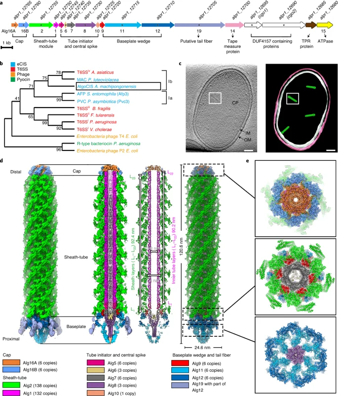
Whole scientific figure with an Algoriphagus cell tomography slice and matching membrane model in panel c, plus gene-cluster, protein-family tree and colourful injection-system molecular reconstructions.
Xu and colleagues’ 2022 Figure 1. Panel c shows a thin tomography slice through an Algoriphagus machipongonensis PR1 cell and its matching model; other panels describe the cell’s AlgoCIS injection system. The molecular structures are not whole bacterial portraits.
Overview
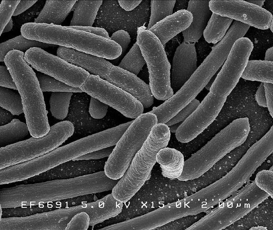
The original taxonomic account describes strain PR1 as nonmotile, non-spore-forming, Gram-negative rods, about 2-3 micrometers long and 0.5 micrometers wide. Colonies were pale pink, with carotenoid pigments. Tested growth was optimal at 30 degrees Celsius and three percent salt; magnesium ions were required. The type strain is PR1, deposited as ATCC BAA-2233 and DSM 24695. It was co-isolated with Salpingoeca rosetta from a Hog Island, Virginia, mud sample. Ribosomal-sequence comparisons placed it near Algoriphagus halophilus and A. lutimaris, while physiological differences helped distinguish it as a species.
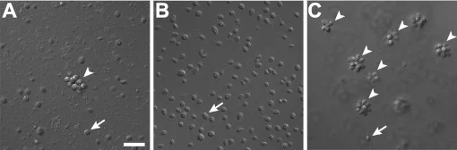
A separate 2012 experimental study by Alegado and colleagues investigated what this bacterium does to its choanoflagellate predator. Of 64 bacterial isolates from the original mixed culture, A. machipongonensis restored rosette formation in a choanoflagellate line that had lost this behavior after antibiotic treatment. Choanoflagellates supplied with this bacterium alone could form colonies. The prey therefore provided a developmental stimulus as well as food, and other tested members of Algoriphagus also had inducing activity. This did not establish that all bacteria, or every strain in the genus, have the same effect.
The active factor RIF-1 was isolated from bacterial lipids and identified as a sulfonolipid. Biological activity occurred in conditioned medium and bacterial cell envelopes; the signal was resistant to treatments that would destroy proteins or nucleic acids. Purified RIF-1 induced rosettes at very low concentrations, but produced fewer colonial cells than an enriched lipid fraction. The investigators proposed that additional molecules or a difference in how the signal reaches the predator could account for this gap. They also considered release in membrane vesicles, without demonstrating that delivery route.
The interaction is especially useful because choanoflagellates are close living relatives of animals. A developmental response in a cultured living relative supplies an experimental model, however, rather than a replay of animal origins. The suggestion that bacterial chemistry influenced early multicellular evolution remains a historical hypothesis. Neither shared isolation nor laboratory induction establishes a permanent intracellular symbiosis: in this account the bacterium is prey and a producer of a signal affecting its predator.
Origins & earliest records
Type strain PR1 was co-isolated from a Hog Island, Virginia, mud sample; the species account appears in IJSEM 63, 2013.
Evidence & interpretation
Taxonomic culture observations and sequence comparisons are distinct from the separately tested rosette-induction mechanism.
Selected bibliography
Documented works and useful reading. This is not a list of every appearance.
- 01
Algoriphagus machipongonensis sp. nov., co-isolated with a colonial choanoflagellate
IJSEM 63:163-168; original species description and comparative tests.
- 02
A bacterial sulfonolipid triggers multicellular development in the closest living relatives of animals
eLife 1:e00013; complete main experimental report. CC Attribution.
Family, evolution & connections
Taxonomic classification
A hierarchy of classified groups, not a chain of direct ancestors.
- domainBacteria
- genusAlgoriphagus
- speciesAlgoriphagus machipongonensis
References
Sources supporting this profile. Linked pages have their own scope and editorial standards.
- ResearchAlegado species descriptionResearch access: 2026-10-05
- ResearchAlegado 2012 RIF-1 experiments, CC AttributionResearch access: 2026-10-05
Image credits
Experimental cell tomography and molecular reconstructions in an original multi-panel figureWhole scientific figure with an Algoriphagus cell tomography slice and matching membrane model in panel c, plus gene-cluster, protein-family tree and colourful injection-system molecular reconstructions.
Xu and colleagues’ 2022 Figure 1. Panel c shows a thin tomography slice through an Algoriphagus machipongonensis PR1 cell and its matching model; other panels describe the cell’s AlgoCIS injection system. The molecular structures are not whole bacterial portraits.