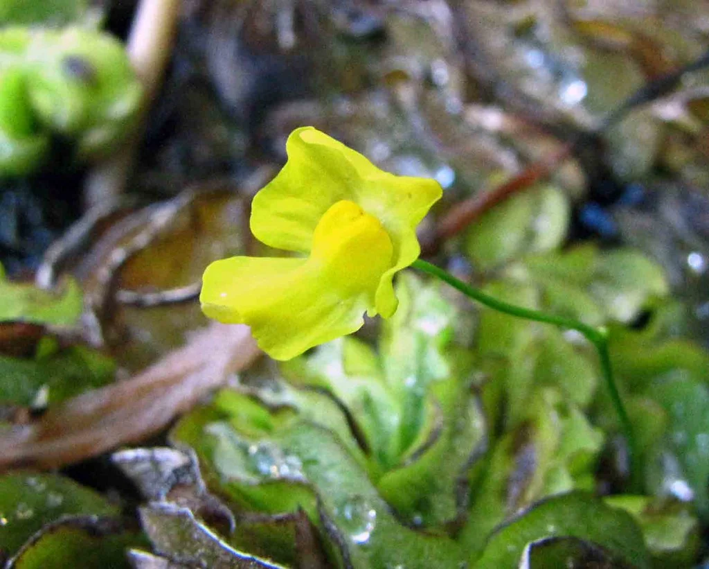
Yellow Utricularia gibba flower photographed by 阿橋 HQ Flower Guide at the Tropical Dream Centre in Okinawa in 2013
This photograph shows a yellow flower identified as Utricularia gibba at Okinawa’s Tropical Dream Centre. It documents a cultivated specimen, not a wild occurrence or the genome-study plant. The underwater bladder traps are not visible in this view.
Overview
Utricularia gibba captures prey through bladder traps that admit water when their pressure-triggered mechanism opens. A 2017 genomic study used Mexican material maintained in sterile tissue culture. Long-read sequencing recovered repetitive regions missed by the earlier assembly, including four complete chromosome contigs; this did not make the entire assembly chromosome-complete.
The published correction gives 29,666 predicted protein-coding genes, replacing the original count of 30,689. Comparative genome structure supports repeated whole-genome duplications alongside localized tandem duplications. Many retained whole-genome duplicates concern regulation, while tandem arrays include candidate digestion, transport and cell-wall functions. Trap-enhanced expression and sequence comparisons identify plausible adaptations; they do not experimentally prove every predicted enzyme function.
The researchers propose an ancient hybridization-related duplication, based on unequal gene loss and expression between reconstructed subgenomes. These are genomic-history inferences, not observed ancestral plants. Their protease tree compares genes, so it should not be presented as a species genealogy.
A separate developmental study by Lee and colleagues in 2019 investigated how the trap acquires its three-dimensional shape. Young traps were approximately spherical; later they became flattened spheroids with a straighter ventral edge. Optical projection tomography and confocal imaging allowed the researchers to compare different sectional planes. Growth tracking began after traps emerged from a tightly curled apex. The estimated initiation time was extrapolated back to a small starting length, rather than directly observed at the moment a trap began.
The imaging conditions matter when interpreting these forms. A primed trap in water has a pressure difference across its wall, whereas the culture-grown traps studied here were closer to a relaxed state. Fixation and preparation for optical tomography could also trigger the trap, altering its outline. The investigators examined these effects and corrected developmental length estimates for measured preparation shrinkage. Their developmental reconstruction therefore concerns quantified shapes under specified conditions, rather than an unchanging outline shared by every trap.
Growth differed between regions and directions. The researchers used these measurements to constrain computational models of a connected tissue sheet. A model combining regional differences in growth with directional growth could account for the overall shape change. Cell and clone patterns and the orientation of internal glandular hairs supplied additional evidence discussed by the authors. They proposed a polarity field comparable to that used in models of planar leaf development, operating here across curved tissue. This is a developmental model, not a directly visualized molecular field or proof that every plant trap evolved through identical changes.
The model deliberately addressed later development after a near-spherical trap had formed. It did not explain the initial curvature, the opening of the mouth, or all the internal folds and thickenings that produce the threshold and door. Nor did it capture every subtle difference in cell size and shape. The study connects measured growth and geometry with hypotheses about developmental control while retaining the distinction between shaping the trap and explaining its complete formation and suction function.
Origins & earliest records
Account based on Lan and colleagues, 2017.
Evidence & interpretation
Long-read assembly, comparative synteny and expression data.
Selected bibliography
Documented works and useful reading. This is not a list of every appearance.
- 01
Long-read sequencing uncovers the adaptive topography of a carnivorous plant genome
PNAS 114, E4435-E4441. Complete main text and main Methods overview read; detailed supplementary Methods not inspected.
- 02
Correction for Lan et al.
PNAS 114, E5483; corrected annotation count 29,666.
- 03
Shaping of a three-dimensional carnivorous trap through modulation of a planar growth mechanism
PLOS Biology 17(10):e3000427, 10 October 2019; DOI 10.1371/journal.pbio.3000427. Complete Introduction, first two growth Results, tissue-model framework, Discussion and material/growth/imaging/quantification/development/triggering Methods read; later model and clonal Results, transformation details, supplements and pixels not fully inspected. Original unrestricted CC Attribution notice; attributed original summary.
Family, evolution & connections
Taxonomic classification
A hierarchy of classified groups, not a chain of direct ancestors.
- GenusUtricularia
- SpeciesUtricularia gibba
References
Sources supporting this profile. Linked pages have their own scope and editorial standards.
- ResearchLan et al. 2017 primary studyResearch access: 2026-10-05
- ResearchPublished annotation correctionResearch access: 2026-10-05
- ResearchShaping of a three-dimensional carnivorous trap through modulation of a planar growth mechanismResearch access: 2026-10-06
Image credits
Scientific plant photographYellow Utricularia gibba flower photographed by 阿橋 HQ Flower Guide at the Tropical Dream Centre in Okinawa in 2013
This photograph shows a yellow flower identified as Utricularia gibba at Okinawa’s Tropical Dream Centre. It documents a cultivated specimen, not a wild occurrence or the genome-study plant. The underwater bladder traps are not visible in this view.