
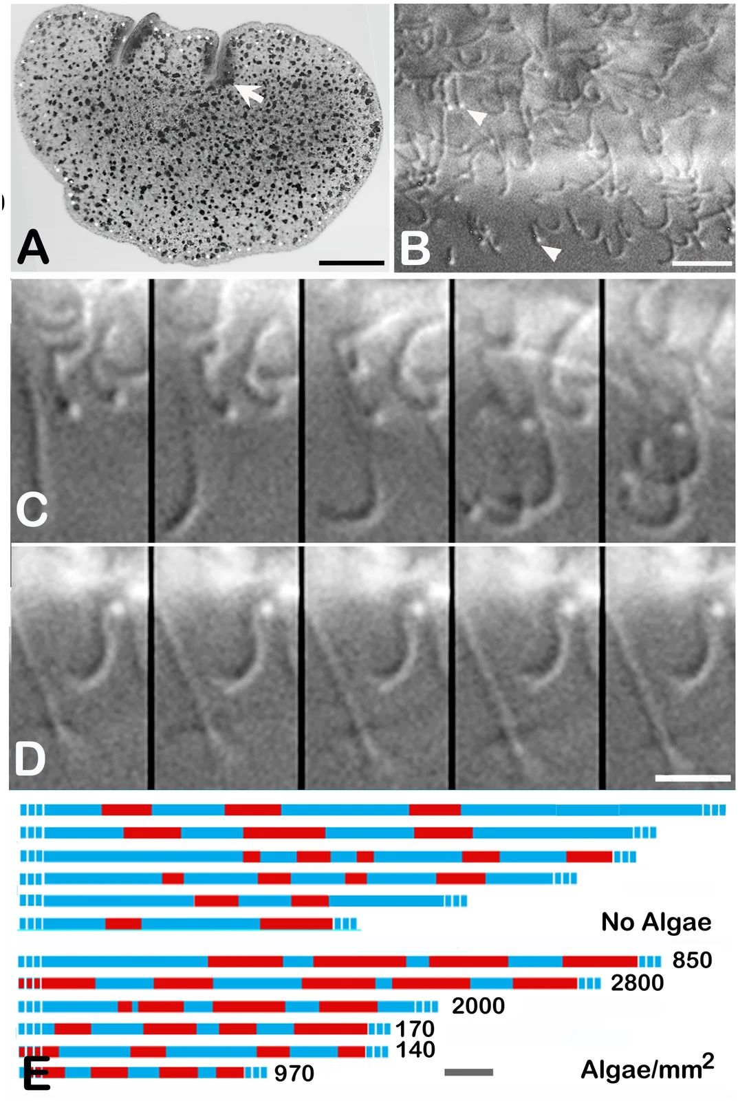
Trichoplax adhaerens Grell-strain whole animal and ciliary microscopy with movement timelines, Smith, Pivovarova and Reese 2015 figure1
Panel A shows a cultured Trichoplax adhaerens of the Grell strain on glass. The other micrographs compare its cilia during movement and pauses; red and blue timelines record those intervals. The visible gray tones depend on transmitted-light and differential-interference-contrast microscopy, while timeline colors encode activity rather than animal coloration.
Overview
Trichoplax adhaerens is a small, flattened marine animal that moves across surfaces using cilia on its underside. It has no internal digestive cavity and no conventional nervous system with synapses. Nevertheless, live microscopy reveals organized changes in movement, secretion and feeding. A 2015 investigation followed these events in the long-maintained Grell laboratory strain, combining fluorescent imaging of living animals with electron microscopy of their cells. Its observations concern this cultured material rather than every placozoan lineage.
While the animal glides, individual cilia beat asynchronously, but their effective strokes point in a common direction. Reorienting those strokes changes the direction of travel. When Trichoplax encounters suitable patches of algae, ciliary beating stops and the animal pauses. Feeding pauses occur much more often in cultures supplied with food than in cultures without it. An animal can resume gliding after digesting a patch and stop again where additional algae remain. These coordinated responses do not require a brain, although the study does not establish a complete mechanism for communication among its cells.
Specialized lipophil cells contain large granules close to the lower body surface. Fluorescent recordings showed those granules being released near underlying algae, followed rapidly by disruption of algal cells. Several lipophils can discharge across a broader region in quick succession. Algae that settled on the animal’s upper surface were not lysed. The resulting digestion occurs outside the animal, in the narrow space between its ventral surface and the substrate. The study identifies a secretory process, rather than demonstrating a named digestive enzyme or a universal response to all possible foods.
During feeding, groups of cells in the central region make elliptical churning movements while the rim remains comparatively still. Material released from the algae disperses beneath the animal and gradually disappears as feeding proceeds. The authors interpret these movements as potentially helping distribute digestive material and support uptake by the ventral surface. Electron microscopy also distinguishes the large secretory granules from other cellular inclusions: their contents and appearance depend on the preparation method, so apparently empty granules in chemically prepared tissue are not evidence that the living cells contain no material.
The observations separate coordinated behavior from its still uncertain signaling machinery. The authors discuss secreted peptides and the possible contributions of internal fiber cells as explanations worth testing; these are hypotheses, not demonstrated neural substitutes. Their comparison with animals and fossil feeding patterns does not make Trichoplax a direct ancestor of other living animals or establish that an Ediacaran fossil fed in the same way. The cultured animal instead offers experimentally accessible evidence that external digestion and organized multicellular behavior can occur without conventional synapses.
Origins & earliest records
Species account centered on Smith, Pivovarova and Reese’s 2015 live-cell and electron-microscopy study of the Grell laboratory strain. No original species-description or type-material examination is claimed.
Evidence & interpretation
Live fluorescent and transmitted-light recordings connect pauses, lipophil secretion and algal lysis; frozen and freeze-substituted electron-microscopy preparations characterize secretory cells. Proposed intercellular signals remain interpretations.
Selected bibliography
Documented works and useful reading. This is not a list of every appearance.
- 01
Coordinated Feeding Behavior in Trichoplax, an Animal without Synapses
PLOS ONE 10(9), e0136098; complete main Results, Discussion, Conclusions and Methods. Grell strain supplied by Leo Buss; the separately examined wild placozoans are not automatically identified as this species. Article explicitly dedicated to CC0.
- 02
The Trichoplax genome and the nature of placozoans
Nature 454:955–960, 21 August 2008; DOI 10.1038/nature07191.
Family, evolution & connections
Selected placozoan lineages in Tessler et al.’s 2022 phylogenomic tree
Seven retained placozoan terminals from Figure 1A. Other placozoan lineages and all external taxa are pruned. H identifiers denote sampled lineages, not invented formal species names.
Scroll sideways to see all branches. With a keyboard, focus the tree and use the arrow keys.
- Selected sampled Placozoa
- Polyplacotoma mediterranea H0
- Remaining sampled Placozoa
- Selected Trichoplax lineages
- Trichoplax H17
- H1 / H2 pair
- Trichoplax H2
- Remaining sampled lineages
- Unnamed placozoan lineage H11
- Selected Cladtertia / Hoilungia lineages
- Cladtertia collaboinventa H23
- Hoilungia hongkongensis H13
IQ-TREE maximum likelihood, C10+GTR20+F+G, on 1,882 orthologs comprising 2,309,771 amino-acid characters. The combined analysis adds 51 MK-model morphological characters weighted 50-fold. The published molecules-only analysis gives the same within-Placozoa branching. The 50-fold morphology weighting is partly subjective and affects the deeper animal root, which is outside this displayed subtree. The source’s mitochondrial comparison instead pairs Trichoplax H1 with H17 rather than H2. Confidence symbols are not converted to exact percentages. Chronogram dates are excluded because no placozoan fossil calibration anchors those nodes; no modern species is shown as a direct ancestor.
Taxonomic classification
A hierarchy of classified groups, not a chain of direct ancestors.
- PhylumPlacozoa
- GenusTrichoplax
- SpeciesTrichoplax adhaerens
Cultured Rhodamonas salina microalgae (study spelling)
Smith, Pivovarova and Reese observed cultured Trichoplax pausing over settled microalgae, stopping ciliary movement and releasing lipophil-cell granules near algal patches. Fluorescence recordings showed algal lysis beneath the animal. These observations describe feeding on laboratory substrates, not an exhaustive natural diet. Proposed chemical or electrical coordination between cells remains an interpretation, rather than a demonstrated synapse-based mechanism.
Located primary passages
Specific passages supporting details in this entry, grouped by their published witness.
The Trichoplax genome and the nature of placozoans
2008
- Conserved regulatory and candidate neural components do not establish conventional nerves or expected functions. Genomics alone cannot determine whether gutless feeding is primitive or derived; phylogenetic placement remains a tested 2008 hypothesis.
Methods, supplements and reanalysis unreviewed. Historical cell-type/species counts are not current facts; homologues do not establish organs or direct ancestry.
References
Sources supporting this profile. Linked pages have their own scope and editorial standards.
- ResearchSmith, Pivovarova and Reese 2015: coordinated feeding, primary study (CC0)Research access: 2026-10-05
Image credits
Scientific organism microscopy and movement figureTrichoplax adhaerens Grell-strain whole animal and ciliary microscopy with movement timelines, Smith, Pivovarova and Reese 2015 figure1
Panel A shows a cultured Trichoplax adhaerens of the Grell strain on glass. The other micrographs compare its cilia during movement and pauses; red and blue timelines record those intervals. The visible gray tones depend on transmitted-light and differential-interference-contrast microscopy, while timeline colors encode activity rather than animal coloration.