
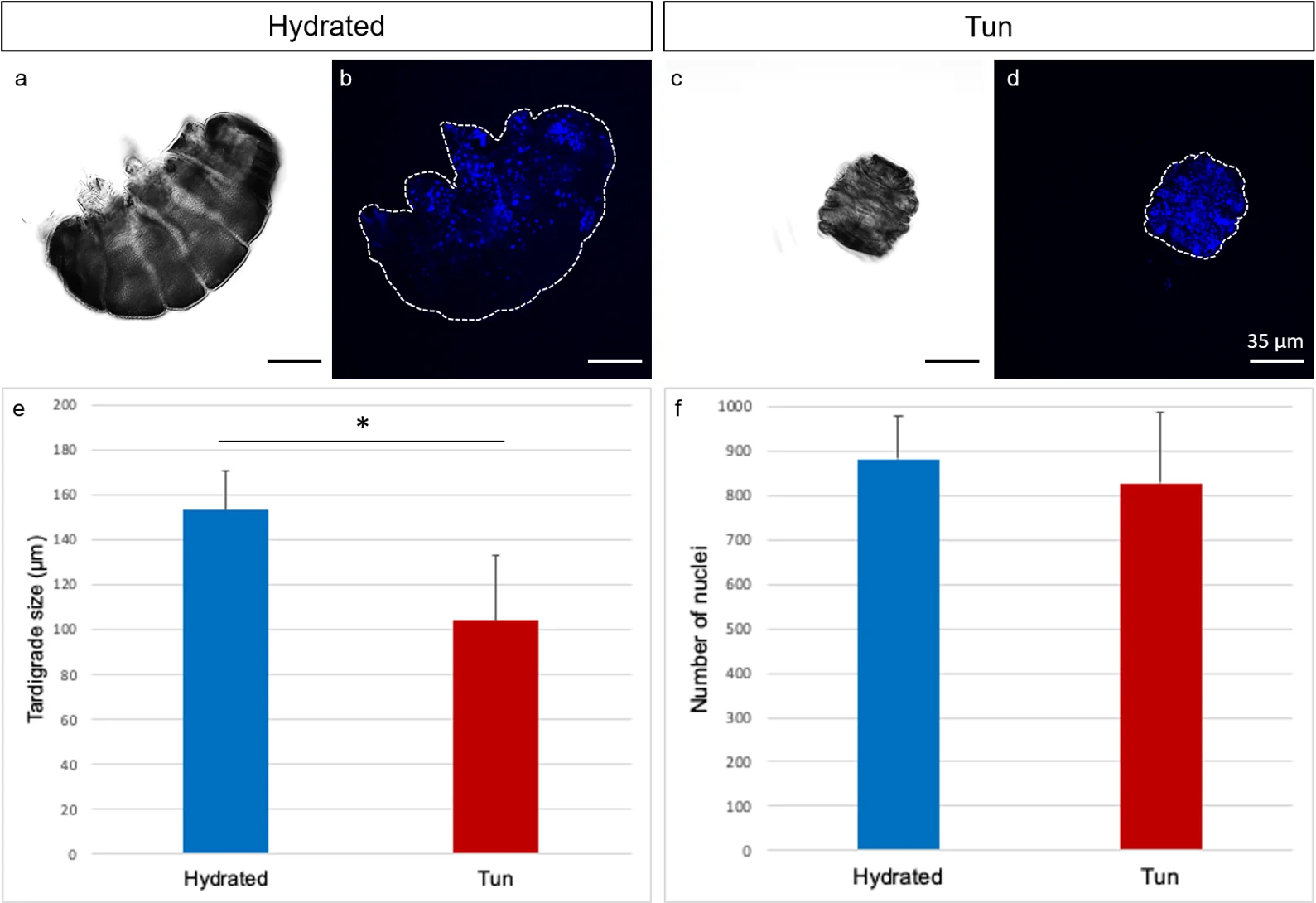
Hydrated and contracted tun forms of Yokozuna-1 Japanese tardigrades in DIC microscopy and blue nuclear staining, with body-size and nucleus-count charts.
Japanese Yokozuna-1 study strain, historically called R. varieornatus in this 2024 paper; the profile uses R. cf. varieornatus. Panels a/c are DIC images; b/d show DAPI-stained nuclei; e/f are comparison charts. No European species portrait substituted.
Overview
The Japanese reference organism called Ramazzottius varieornatus in the 2019 study is a tardigrade, one of the small invertebrates commonly called water bears. Tardigrades have four pairs of claw-bearing legs; active terrestrial forms depend on a surrounding film of water. Some can survive drying by entering anhydrobiosis, a dormant dehydrated state from which activity resumes after rehydration. This physiological transition is distinct from simply remaining active in extreme conditions. A 2019 biochemical study uses this species to investigate one molecular contributor to protection against DNA damage.
The study examines Dsup, short for damage suppressor, a highly charged nuclear protein with extensive disordered regions. Rather than testing only naked DNA, Chavez and colleagues reconstruct nucleosomes: DNA wrapped around histone proteins, the fundamental units of chromatin. Purified recombinant Dsup binds these structures more readily than corresponding free DNA. Experiments using two different positioning sequences support a preference for nucleosomes rather than a single particular DNA sequence. Dsup also enters experimentally assembled arrays of nucleosomes without substantially disrupting their regular organization.
Protection is tested in a defined chemical system that generates hydroxyl radicals, reactive molecules capable of fragmenting DNA. Adding Dsup leaves more intact DNA after the treatment, with stronger protection of chromatin than free DNA under the studied conditions. A comparison protein, human TFIIB, does not show detectable protection in the same assay. The authors propose that disordered regions of nucleosome-bound Dsup cover and shield DNA. The protective effect is measured; the precise physical arrangement of that covering remains a mechanistic model.
Changing the protein helps connect binding with protection. Removing its C-terminal region greatly reduces nucleosome binding and protective activity. Three amino-acid substitutions in a smaller conserved region also impair both functions. That region resembles part of vertebrate HMGN proteins, another family associated with nucleosomes. Sequence resemblance does not establish that tardigrades acquired a vertebrate gene: the authors leave its evolutionary origin unresolved. Histone H1 and Dsup can bind the same nucleosome simultaneously, showing that their presence need not be mutually exclusive.
The comparison includes a corresponding protein from Hypsibius exemplaris. Similar neighboring genes, sequence features and biochemical activity support the authors’ identification of the two proteins as orthologs. This extends the molecular result beyond a single tardigrade species, without showing that every tardigrade uses the same mechanism. The 2019 experiments use purified components, including histones from another animal; they do not reproduce the complete living tardigrade. A contribution to survival during dehydration is plausible, while medical benefit or increased human lifespan is not demonstrated by these assays.
Identification requires a strain qualification. Emdee and colleagues’ 2024 primary study explicitly distinguishes its Danish R. varieornatus population from the Japanese population underlying the public reference genome, treating the latter as a different species. The Dsup results here remain attached to the Japanese reference material and the name used in the 2019 paper. They should not be silently transferred to European R. varieornatus; this profile does not propose a new formal species name.
Origins & earliest records
Chavez and colleagues’ 2019 biochemical investigation compares recombinant proteins encoded by R. varieornatus and Hypsibius exemplaris. The R. varieornatus genomic comparison uses strain YOKOZUNA-1; this is research material, not a nomenclatural type designation.
Evidence & interpretation
Purified-protein binding assays, reconstructed chromatin, chemically induced DNA cleavage and altered Dsup proteins connect nucleosome association with protection. Whole-animal dehydration survival and clinical benefit are not measured by this study.
Selected bibliography
Documented works and useful reading. This is not a list of every appearance.
- 01
The tardigrade damage suppressor protein binds to nucleosomes and protects DNA from hydroxyl radicals
eLife 8:e47682; complete main Results, Discussion and Materials and methods. Original adaptation of CC BY 4.0 article, https://creativecommons.org/licenses/by/4.0/.
- 02
Osmotic stress tolerance and transcriptomic response of Ramazzottius varieornatus (Eutardigrada: Ramazzottiidae) following tun formation
Zoological Journal of the Linnean Society 200,220–229; selected complete Differential expression analyses Methods paragraph explicitly distinguishes Danish and Japanese populations. Online 4 July 2023; issue 2024. No full osmobiosis account claimed here.
- 03
Stress-dependent cell stiffening by tardigrade tolerance proteins that reversibly form a filamentous network and gel
Tanaka and coauthors, PLOS Biology20(9)e3001780,6September2022; DOI10.1371/journal.pbio.3001780
- 04
Extremotolerant tardigrade genome and improved radiotolerance of human cultured cells by tardigrade-unique protein
Nature Communications7:12808,20September2016.
Family, evolution & connections
Taxonomic classification
A hierarchy of classified groups, not a chain of direct ancestors.
- PhylumTardigrada
- GenusRamazzottius
Engineered HEK293 cells
YOKOZUNA-1 Dsup reduced irradiation-associated DNA damage in selected cultured cells; whole-human protection was not established.
Located primary passages
Specific passages supporting details in this entry, grouped by their published witness.
Stress-dependent cell stiffening by tardigrade tolerance proteins that reversibly form a filamentous network and gel
Tanaka and coauthors, PLOS Biology20(9)e3001780,6September2022; DOI10.1371/journal.pbio.3001780
- Proteins isolated from this tardigrade form reversible networks or granules under dehydration-like stress. Experiments in cultured human and insect cells and artificial droplets show that CAHS3 can increase stiffness and help resist shrinkage. These model systems support a proposed protective mechanism; the authors do not establish that filament formation alone explains survival or directly demonstrate the same networks inside a drying tardigrade.
Full Methods, supplementary experiments, movies and raw datasets not independently reviewed or replicated. Desolvating-agent effects and protein-concentration estimates are qualified by the authors. Different witness from the2019 Dsup paper. The article calls the Japanese study organism R.varieornatus; this supplement preserves that historical identification and does not independently confirm species identity. The current profile qualifies it as R.cf.varieornatus study strain.
Primary source witness
2016 report.
- In YOKOZUNA-1, historically identified as R.varieornatus, the study identified Dsup and showed reduced radiation-associated DNA damage in human cultured cells. Knockdown removed protection. The authors distinguish damage suppression from faster repair and warn that cultured-cell adaptation may contribute; whole-animal tolerance requires additional factors.
Other Results/Methods, supplements and raw analyses uninspected. Later Japanese/Danish taxonomic distinction preserved; no human-treatment inference.
References
Sources supporting this profile. Linked pages have their own scope and editorial standards.
- ResearchChavez et al. 2019 primary biochemical studyResearch access: 2026-10-05
- ResearchEmdee et al. 2024 primary population qualificationResearch access: 2026-10-05
Image credits
Whole experimental tardigrade microscopy comparison figureHydrated and contracted tun forms of Yokozuna-1 Japanese tardigrades in DIC microscopy and blue nuclear staining, with body-size and nucleus-count charts.
Japanese Yokozuna-1 study strain, historically called R. varieornatus in this 2024 paper; the profile uses R. cf. varieornatus. Panels a/c are DIC images; b/d show DAPI-stained nuclei; e/f are comparison charts. No European species portrait substituted.