
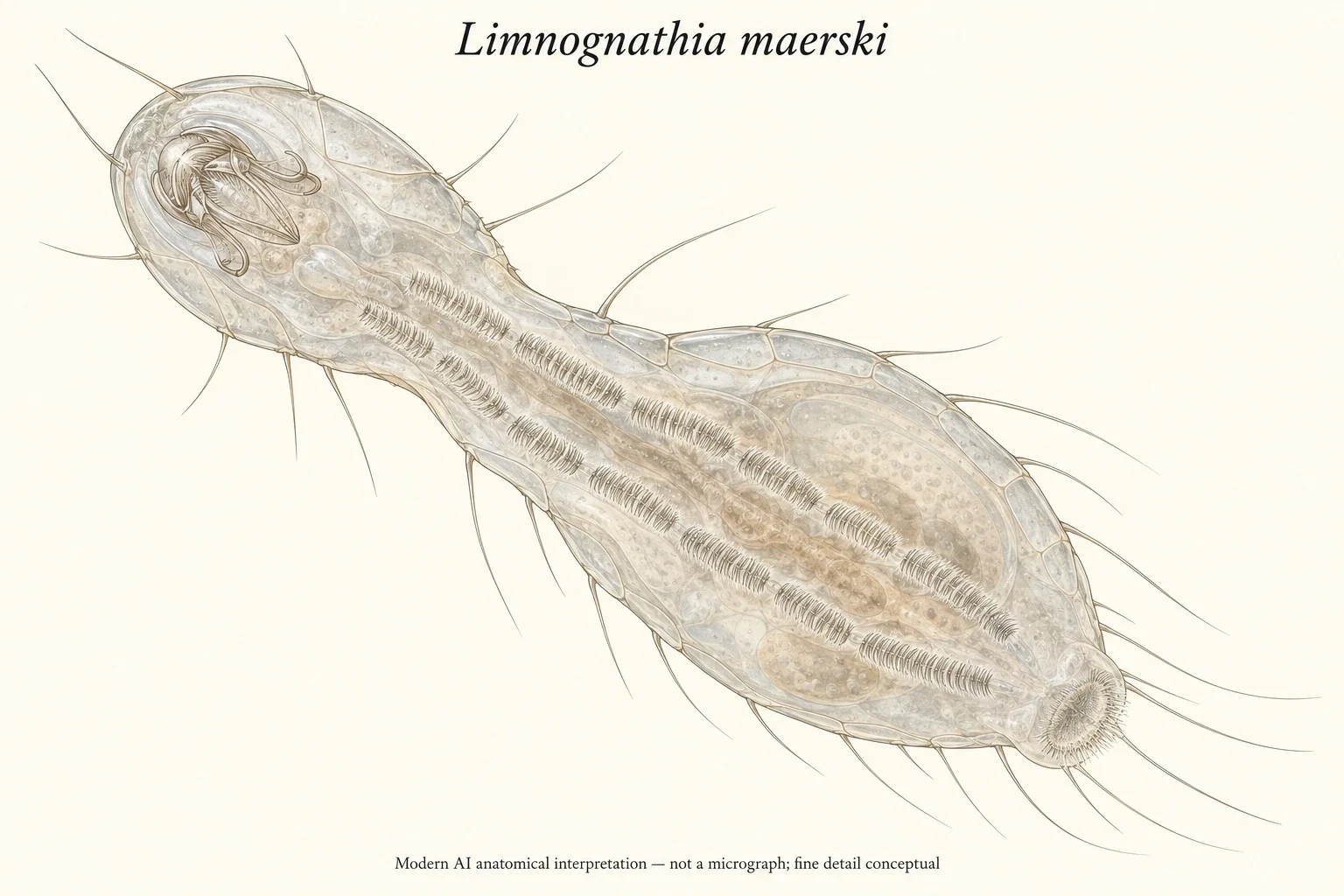
AI micrognathozoan anatomy; fine detail conceptual.
Modern AI interpretation: internal jaws, ciliary rows and adhesive pad; not a micrograph.
Overview
The head, thorax and abdomen bear dorsal and lateral epidermal plates, but the ventral epidermis lacks internal plates. Two ventral ciliary rows provide locomotion; a posterior adhesive ciliated pad assists contact. Tactile sensoria project from the body, while complex jaws lie inside the pharynx.
A published correction to the original journal cover is essential to scale and colour. Its pictured animal measured 128 micrometres, not 128 centimetres. The correction also states that the vivid colour in the Nomarski photomicrograph was an optical effect, while the living animal is transparent. Neither a giant worm nor a naturally rainbow-coloured body is justified by that image.
Jaw similarities suggested Gnathifera ancestry to the 2000 authors. Their particular sister-group proposal remains historical, not a newly computed tree.
Recent species delimitation changes the older account of a single widespread species. Sato and coauthors combined anatomy and single-specimen transcriptomic data across Greenland, the Pyrenees and a Subantarctic population. They recognised the Subantarctic population as Limnognathia desmeti but did not definitively split the Greenland and Pyrenean samples. Visible similarity therefore cannot safely assign an unidentified Crozet animal to L. maerski.
Origins & earliest records
Kristensen and Funch described the West Greenland animal in 2000. Only adult females occurred in the original sample.
Evidence & interpretation
Cited text only; no sourced image reused.
Selected bibliography
Documented works and useful reading. This is not a list of every appearance.
- 01
- 02
- 03
Family, evolution & connections
Taxonomic classification
A hierarchy of classified groups, not a chain of direct ancestors.
Source note: The 2025 study uses phylum rank; L. desmeti remains separate.
- PhylumMicrognathozoa
- GenusLimnognathia
- SpeciesLimnognathia maerski
References
Sources supporting this profile. Linked pages have their own scope and editorial standards.
- Primary textSource recordResearch access: 2026-10-11
- Primary textSource recordResearch access: 2026-10-11
- Primary textSource recordResearch access: 2026-10-11
Image credits
AI-generated interpretationAI micrognathozoan anatomy; fine detail conceptual.
Modern AI interpretation: internal jaws, ciliary rows and adhesive pad; not a micrograph.