
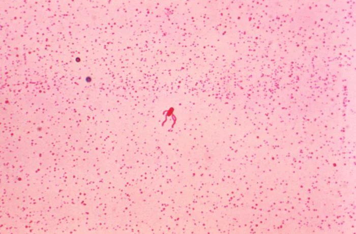
Pink field containing a small stained rod-shaped bacterium near the centre, with several fine flagella visible around it.
CDC identifies the central specimen as Bacillus subtilis; the stain reveals peritrichous flagella. Pink coloration comes from the stained preparation. Original supplied low-resolution image retained (700x460); no magnification or scale inferred. CDC supplies the image freely; use does not imply agency endorsement.
Overview
Kunst and colleagues reported in 1997 that laboratory strain 168 has a genome of 4,214,810 base pairs with roughly 4,100 protein-coding genes. These figures describe that strain, not every isolate.
The same study noted expanded gene families and at least ten prophage or prophage-like elements, and presented the sequence as a link between molecular genetics and cell physiology.
Sporulation starts when phosphorylated Spo0A triggers asymmetric division into a mother cell and a forespore, which the mother cell then engulfs. Four alternative sigma factors stage gene expression across the two compartments. A comparative-genomics study described a conserved core of about 100 sporulation genes and showed ytaF, ylmC and ylzA are needed for efficient sporulation.
Origins & earliest records
First description and naming history are unverified. How sporulation arose is a phylogenetic hypothesis, not a directly observed event.
Evidence & interpretation
Genome figures rest on the indexed 1997 Nature abstract and indexed author-hosted PDF; publisher full text was not opened. Sporulation details come from an opened 2013 ASM article.
Selected bibliography
Documented works and useful reading. This is not a list of every appearance.
- 01
The complete genome sequence of the Gram-positive bacterium Bacillus subtilis
Selected bibliography; strain 168 genome paper, Nature 390:249–256.
- 02
A Genomic Signature and the Identification of New Sporulation Genes
Selected bibliography; comparative sporulation-gene study.
Family, evolution & connections
Taxonomic classification
A hierarchy of classified groups, not a chain of direct ancestors.
Source note: Ranked groups in the NCBI Taxonomy snapshot accessed 7 October 2026; these are not identified direct ancestors. NCBI uses Caryophanales and Bacillota; older classifications may use Bacillales and Firmicutes. LPSN confirms the species name and notes that some historical bioindicator strains were reassigned to Bacillus atrophaeus.
- domainBacteria
- kingdomBacillati
- phylumBacillota
- classBacilli
- orderCaryophanales
- familyBacillaceae
- genusBacillus
- speciesBacillus subtilis
References
Sources supporting this profile. Linked pages have their own scope and editorial standards.
- Scientific paper (indexed abstract and author PDF)The complete genome sequence of the Gram-positive bacterium Bacillus subtilisResearch access: 2026-10-06
- Scientific paperA Genomic Signature and the Identification of New Sporulation GenesResearch access: 2026-10-06
- Scientific classification referenceNCBI Taxonomy: Bacillus subtilisResearch access: 2026-10-07
- Scientific classification referenceLPSN: Bacillus subtilisResearch access: 2026-10-07
Image credits
Leifson flagella-stained light micrographPink field containing a small stained rod-shaped bacterium near the centre, with several fine flagella visible around it.
CDC identifies the central specimen as Bacillus subtilis; the stain reveals peritrichous flagella. Pink coloration comes from the stained preparation. Original supplied low-resolution image retained (700x460); no magnification or scale inferred. CDC supplies the image freely; use does not imply agency endorsement.